 Movie
Movie Controller

[English] 日本語
 Yorodumi
- PDB-9rjs: Structure of the Bacteriophage PhiKZ non-virion RNA Polymerase bo... -
+ Open data

- Basic information
| Entry | Database: PDB / ID: 9rjs | ||||||||||||||||||||||||||||||
|---|---|---|---|---|---|---|---|---|---|---|---|---|---|---|---|---|---|---|---|---|---|---|---|---|---|---|---|---|---|---|---|
| Title | Structure of the Bacteriophage PhiKZ non-virion RNA Polymerase bound to an analogue of its promoter | ||||||||||||||||||||||||||||||
 Components Components |
| ||||||||||||||||||||||||||||||
 Keywords Keywords | RNA BINDING PROTEIN / PhiKZ / nvRNAP / RNA / DNA / transcription | ||||||||||||||||||||||||||||||
| Function / homology | metal ion binding / DNA / DNA (> 10) / DNA-directed RNA polymerase / PHIKZ056.1 / PHIKZ123 / PHIKZ074 / PHIKZ068 Function and homology information Function and homology information | ||||||||||||||||||||||||||||||
| Biological species |  Phikzvirus phiKZ Phikzvirus phiKZ | ||||||||||||||||||||||||||||||
| Method | ELECTRON MICROSCOPY / single particle reconstruction / cryo EM / Resolution: 2.59 Å | ||||||||||||||||||||||||||||||
 Authors Authors | Chen, C. / de Martin Garrido, N. / Yakunina, M. / Aylett, C.H.S. | ||||||||||||||||||||||||||||||
| Funding support |  United Kingdom, 2items United Kingdom, 2items
| ||||||||||||||||||||||||||||||
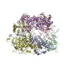
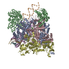
 Citation Citation | Journal: IUCrJ / Year: 2026 Title: Structure of the bacteriophage PhiKZ non-virion RNA polymerase bound to a p119L open promoter analogue. Authors: Chao Sheng Chen / Natàlia de Martín Garrido / Maria Yakunina / Christopher H S Aylett /  Abstract: Bacteriophage ΦKZ (PhiKZ) was the first identified member of a family of massive bacterial viruses. ΦKZ infects Pseudomonas aeruginosa, which kills tens of thousands every year, and it therefore ...Bacteriophage ΦKZ (PhiKZ) was the first identified member of a family of massive bacterial viruses. ΦKZ infects Pseudomonas aeruginosa, which kills tens of thousands every year, and it therefore has potential as a bacteriophage therapy. On infection, ΦKZ forms a `nucleus' to protect its genome by excluding host immune systems. This barrier means that it has had to become independent of the host transcriptional apparatus; it cannot simply recruit the host RNA polymerase (RNAP) to its promoters as it is excluded from the viral DNA, and therefore it expresses and imports its own non-virion RNA polymerase (nvRNAP). The ΦKZ nvRNAP, and related jumbo-phage RNAPs including that from bacteriophage AR9, are particularly noteworthy. Unlike typical viral RNAPs which are formed as only a single subunit, it is a non-canonical multi-subunit RNAP directly related to those from eubacteria, and more distantly eukaryotes and archaea. It encompasses four proteins representing patchwork homologues of the eubacterial β/β' subunits, and a fifth that appears to have evolved from a σ factor, but no homologues of the α or ω subunits required for formation of a catalytically active complex in eubacterial RNAPs. Its mechanism of promoter recognition is also highly divergent; transcription is initiated from a site marked only by a tiny four-base consensus sequence co-located with the start site. We have resolved the structure of the ΦKZ nvRNAP bound to an open analogue of its cognate promoter, p119L, revealing that while the σ-factor-like subunit GP68 is involved in bubble stabilization, the sequence-specific promoter consensus sequence is bound between the lobe of the β-subunit homologue GP123 and the enzymatic core of the complex. Our results shed light on the differences between mechanisms of promoter recognition in the ΦKZ nvRNAP and canonical eubacterial RNAPs, and on the uniquely specialized features of bacteriophage transcriptional apparatuses in general. | ||||||||||||||||||||||||||||||
| History |
|
- Structure visualization
| Structure viewer | Molecule: MolmilJmol/JSmol |
|---|
- Downloads & links
-Download
| PDBx/mmCIF format | 9rjs.cif.gz | 671.4 KB | Display | PDBx/mmCIF format |
|---|---|---|---|---|
| PDB format | pdb9rjs.ent.gz | 427.5 KB | Display | PDB format |
| PDBx/mmJSON format | 9rjs.json.gz | Tree view | PDBx/mmJSON format | |
| Others |  Other downloads Other downloads |
-Validation report
| Arichive directory | https://data.pdbj.org/pub/pdb/validation_reports/rj/9rjsftp://data.pdbj.org/pub/pdb/validation_reports/rj/9rjs | HTTPS FTP |
|---|
-Related structure data
| Related structure data |  54012MC M: map data used to model this data C: citing same article ( |
|---|---|
| Similar structure data |

-Links
 PDBj
PDBj


- Assembly
| Deposited unit | 
|
|---|---|
| 1 |
|
-Components
-Protein , 5 types, 5 molecules ABCDE
| #1: Protein | Mass: 57976.605 Da / Num. of mol.: 1 Source method: isolated from a genetically manipulated source Source: (gene. exp.) Phikzvirus phiKZ / Production host:  |
|---|---|
| #2: Protein | Mass: 59442.824 Da / Num. of mol.: 1 Source method: isolated from a genetically manipulated source Details: Numbering correct in original mmCIF file / Source: (gene. exp.) Phikzvirus phiKZ / Production host: |
| #3: Protein | Mass: 78780.453 Da / Num. of mol.: 1 Source method: isolated from a genetically manipulated source Source: (gene. exp.) Phikzvirus phiKZ / Production host: |
| #4: Protein | Mass: 77513.461 Da / Num. of mol.: 1 Source method: isolated from a genetically manipulated source Source: (gene. exp.) Phikzvirus phiKZ / Production host: |
| #5: Protein | Mass: 62959.090 Da / Num. of mol.: 1 Source method: isolated from a genetically manipulated source Source: (gene. exp.) Phikzvirus phiKZ / Production host: |
-DNA chain , 2 types, 2 molecules XY
| #6: DNA chain | Mass: 23218.951 Da / Num. of mol.: 1 / Source method: obtained synthetically / Source: (synth.) Phikzvirus phiKZ |
|---|---|
| #7: DNA chain | Mass: 22881.719 Da / Num. of mol.: 1 / Source method: obtained synthetically / Source: (synth.) Phikzvirus phiKZ |
-Non-polymers , 1 types, 2 molecules 
| #8: Chemical |  Mass: 65.409 Da / Num. of mol.: 2 / Source method: obtained synthetically / Formula: Zn Mass: 65.409 Da / Num. of mol.: 2 / Source method: obtained synthetically / Formula: Zn |
|---|
-Details
| Has ligand of interest | N |
|---|---|
| Has protein modification | N |
-Experimental details
-Experiment
| Experiment | Method: ELECTRON MICROSCOPY |
|---|---|
| EM experiment | Aggregation state: PARTICLE / 3D reconstruction method: single particle reconstruction |
- Sample preparation
| Component | Name: PhiKZ non-virion RNA polymerase bound to an analogue of promoter p119L Type: COMPLEX Details: Mismatches introduced around the transcription start site to stably open the bubble Entity ID: #1-#7 / Source: RECOMBINANT | ||||||||||||||||||||
|---|---|---|---|---|---|---|---|---|---|---|---|---|---|---|---|---|---|---|---|---|---|
| Molecular weight | Value: 0.4 MDa / Experimental value: NO | ||||||||||||||||||||
| Source (natural) | Organism: Pseudomonas phage phiKZ (virus) | ||||||||||||||||||||
| Source (recombinant) | Organism: | ||||||||||||||||||||
| Buffer solution | pH: 8 | ||||||||||||||||||||
| Buffer component |
| ||||||||||||||||||||
| Specimen | Conc.: 0.1 mg/ml / Embedding applied: NO / Shadowing applied: NO / Staining applied: NO / Vitrification applied: YES Details: PhiKZ nvRNAP complexes were mixed at 0.1 mg/mL final concentration with the P119L promoter analogue at a 1:1 ratio. Reactions were incubated for 30 min at 25 C before use. | ||||||||||||||||||||
| Specimen support | Grid material: GOLD / Grid mesh size: 300 divisions/in. / Grid type: Quantifoil R2/1 | ||||||||||||||||||||
| Vitrification | Instrument: FEI VITROBOT MARK IV / Cryogen name: ETHANE / Humidity: 100 % / Chamber temperature: 289 K Details: Settings: -4 blot force, 4 s waiting time and 0.5-1 s blotting time. |
- Electron microscopy imaging
| Experimental equipment |  Model: Titan Krios / Image courtesy: FEI Company |
|---|---|
| Microscopy | Model: TFS KRIOS |
| Electron gun | Electron source:  FIELD EMISSION GUN / Accelerating voltage: 300 kV / Illumination mode: FLOOD BEAM FIELD EMISSION GUN / Accelerating voltage: 300 kV / Illumination mode: FLOOD BEAM |
| Electron lens | Mode: BRIGHT FIELD / Nominal magnification: 130000 X / Nominal defocus max: 2500 nm / Nominal defocus min: 750 nm / Cs: 2.7 mm / C2 aperture diameter: 50 µm / Alignment procedure: ZEMLIN TABLEAU |
| Specimen holder | Cryogen: NITROGEN / Specimen holder model: FEI TITAN KRIOS AUTOGRID HOLDER |
| Image recording | Electron dose: 50 e/Å2 / Film or detector model: GATAN K3 BIOQUANTUM (6k x 4k) / Num. of grids imaged: 1 / Num. of real images: 20670 / Details: Images collected as TIFF movies |
| EM imaging optics | Energyfilter name: GIF Bioquantum / Energyfilter slit width: 10 eV |
| Image scans | Sampling size: 5 µm / Width: 5760 / Height: 4092 |
- Processing
| EM software |
| ||||||||||||||||||||||||||||||||||||||||||||||||||||||
|---|---|---|---|---|---|---|---|---|---|---|---|---|---|---|---|---|---|---|---|---|---|---|---|---|---|---|---|---|---|---|---|---|---|---|---|---|---|---|---|---|---|---|---|---|---|---|---|---|---|---|---|---|---|---|---|
| CTF correction | Type: PHASE FLIPPING AND AMPLITUDE CORRECTION | ||||||||||||||||||||||||||||||||||||||||||||||||||||||
| Particle selection | Num. of particles selected: 4178816 | ||||||||||||||||||||||||||||||||||||||||||||||||||||||
| Symmetry | Point symmetry: C1 (asymmetric) | ||||||||||||||||||||||||||||||||||||||||||||||||||||||
| 3D reconstruction | Resolution: 2.59 Å / Resolution method: FSC 0.143 CUT-OFF / Num. of particles: 329400 / Algorithm: FOURIER SPACE / Num. of class averages: 1 / Symmetry type: POINT | ||||||||||||||||||||||||||||||||||||||||||||||||||||||
| Atomic model building | B value: 104.82 / Protocol: AB INITIO MODEL / Space: REAL / Target criteria: CC | ||||||||||||||||||||||||||||||||||||||||||||||||||||||
| Atomic model building | 3D fitting-ID: 1
| ||||||||||||||||||||||||||||||||||||||||||||||||||||||
| Refinement | Cross valid method: NONE Stereochemistry target values: GeoStd + Monomer Library + CDL v1.2 | ||||||||||||||||||||||||||||||||||||||||||||||||||||||
| Displacement parameters | Biso mean: 101.01 Å2 | ||||||||||||||||||||||||||||||||||||||||||||||||||||||
| Refine LS restraints |
|
