 Movie
Movie Controller

[English] 日本語
 Yorodumi
- PDB-9raj: Class A CTX-M-14 E166A mutant in complex with Dicloxacillin at ro... -
+ Open data

- Basic information
| Entry | Database: PDB / ID: 9raj | |||||||||
|---|---|---|---|---|---|---|---|---|---|---|
| Title | Class A CTX-M-14 E166A mutant in complex with Dicloxacillin at room temperature | |||||||||
 Components Components | Beta-lactamase | |||||||||
 Keywords Keywords | PROTEIN BINDING / Antibiotic resistance / Class A beta-lactamase / isoxazolyl penicillins | |||||||||
| Function / homology |  Function and homology information Function and homology informationbeta-lactam antibiotic catabolic process / beta-lactamase activity / beta-lactamase / response to antibiotic Similarity search - Function | |||||||||
| Biological species |  Klebsiella pneumoniae (bacteria) Klebsiella pneumoniae (bacteria) | |||||||||
| Method |  X-RAY DIFFRACTION / SYNCHROTRON / MOLECULAR REPLACEMENT / Resolution: 1.7 Å X-RAY DIFFRACTION / SYNCHROTRON / MOLECULAR REPLACEMENT / Resolution: 1.7 Å | |||||||||
 Authors Authors | Gore, G. / Schulz, E.C. | |||||||||
| Funding support |  Germany, 2items Germany, 2items
| |||||||||
 Citation Citation | Journal: Commun Chem / Year: 2025 Title: Binding mode of Isoxazolyl Penicillins to a Class-A beta-lactamase at ambient conditions. Authors: Gore, G. / Prester, A. / von Stetten, D. / Bartels, K. / Schulz, E.C. | |||||||||
| History |
|
- Structure visualization
| Structure viewer | Molecule: MolmilJmol/JSmol |
|---|
- Downloads & links
-Download
| PDBx/mmCIF format | 9raj.cif.gz | 89.8 KB | Display | PDBx/mmCIF format |
|---|---|---|---|---|
| PDB format | pdb9raj.ent.gz | 54 KB | Display | PDB format |
| PDBx/mmJSON format | 9raj.json.gz | Tree view | PDBx/mmJSON format | |
| Others |  Other downloads Other downloads |
-Validation report
| Arichive directory | https://data.pdbj.org/pub/pdb/validation_reports/ra/9rajftp://data.pdbj.org/pub/pdb/validation_reports/ra/9raj | HTTPS FTP |
|---|
-Related structure data


-Links
 PDBj
PDBj


- Assembly
| Deposited unit | 
| ||||||||||||
|---|---|---|---|---|---|---|---|---|---|---|---|---|---|
| 1 |
| ||||||||||||
| Unit cell |
| ||||||||||||
| Components on special symmetry positions |
|
-Components
| #1: Protein | Mass: 30951.256 Da / Num. of mol.: 1 / Mutation: E166A Source method: isolated from a genetically manipulated source Source: (gene. exp.) Klebsiella pneumoniae (bacteria) / Gene: KPHS_p301310Production host: References: UniProt: A0A0H3H219, beta-lactamase |
|---|---|
| #2: Chemical | ChemComp-DXU / 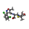  Mass: 478.390 Da / Num. of mol.: 1 / Source method: obtained synthetically / Formula: C19H25Cl2N3O5S / Feature type: SUBJECT OF INVESTIGATION Mass: 478.390 Da / Num. of mol.: 1 / Source method: obtained synthetically / Formula: C19H25Cl2N3O5S / Feature type: SUBJECT OF INVESTIGATION |
| #3: Water | ChemComp-HOH /  Mass: 18.015 Da / Num. of mol.: 172 / Source method: isolated from a natural source / Formula: H2O Mass: 18.015 Da / Num. of mol.: 172 / Source method: isolated from a natural source / Formula: H2O |
| Has ligand of interest | Y |
| Has protein modification | Y |
-Experimental details
-Experiment
| Experiment | Method: X-RAY DIFFRACTION / Number of used crystals: 1 |
|---|
- Sample preparation
| Crystal | Density Matthews: 1.94 Å3/Da / Density % sol: 36.49 % |
|---|---|
| Crystal grow | Temperature: 293 K / Method: batch mode Details: CTX-M-14 solution (22 mg/ml) was mixed with 45% precipitant solution (40% PEG8000, 200mM lithium sulfate, 100mM sodium acetate, pH 4.5) and with 5% undiluted seed stock in batch crystallization setups |
-Data collection
| Diffraction | Mean temperature: 293 K / Serial crystal experiment: Y |
|---|---|
| Diffraction source | Source: SYNCHROTRON / Site: PETRA III, EMBL c/o DESY / Beamline: P14 (MX2) / Wavelength: 0.976 Å |
| Detector | Type: DECTRIS EIGER R 4M / Detector: PIXEL / Date: Sep 23, 2021 |
| Radiation | Protocol: SINGLE WAVELENGTH / Monochromatic (M) / Laue (L): M / Scattering type: x-ray |
| Radiation wavelength | Wavelength: 0.976 Å / Relative weight: 1 |
| Reflection | Resolution: 1.7→117.65 Å / Num. obs: 27982 / % possible obs: 100 % / Redundancy: 92.8 % / Biso Wilson estimate: 21.03 Å2 / CC1/2: 0.938 / CC star: 0.984 / Net I/σ(I): 4.04 |
| Reflection shell | Resolution: 1.7→1.76 Å / Num. unique obs: 2734 / CC1/2: 0.535 / CC star: 0.835 |
| Serial crystallography sample delivery | Method: fixed target |
| Serial crystallography sample delivery fixed target | Sample holding: Hare-Chip , silicon |
- Processing
| Software |
| |||||||||||||||||||||||||||||||||||||||||||||||||||||||||||||||||||||||||||||
|---|---|---|---|---|---|---|---|---|---|---|---|---|---|---|---|---|---|---|---|---|---|---|---|---|---|---|---|---|---|---|---|---|---|---|---|---|---|---|---|---|---|---|---|---|---|---|---|---|---|---|---|---|---|---|---|---|---|---|---|---|---|---|---|---|---|---|---|---|---|---|---|---|---|---|---|---|---|---|
| Refinement | Method to determine structure: MOLECULAR REPLACEMENT / Resolution: 1.7→78.07 Å / SU ML: 0.2179 / Cross valid method: FREE R-VALUE / σ(F): 1.35 / Phase error: 23.9346 Stereochemistry target values: GeoStd + Monomer Library + CDL v1.2
| |||||||||||||||||||||||||||||||||||||||||||||||||||||||||||||||||||||||||||||
| Solvent computation | Shrinkage radii: 0.9 Å / VDW probe radii: 1.1 Å / Solvent model: FLAT BULK SOLVENT MODEL | |||||||||||||||||||||||||||||||||||||||||||||||||||||||||||||||||||||||||||||
| Displacement parameters | Biso mean: 24.9 Å2 | |||||||||||||||||||||||||||||||||||||||||||||||||||||||||||||||||||||||||||||
| Refinement step | Cycle: LAST / Resolution: 1.7→78.07 Å
| |||||||||||||||||||||||||||||||||||||||||||||||||||||||||||||||||||||||||||||
| Refine LS restraints |
| |||||||||||||||||||||||||||||||||||||||||||||||||||||||||||||||||||||||||||||
| LS refinement shell |
|
