regulation of response to type II interferon / NAD+-protein-C-terminal glycine ADP-ribosyltransferase activity / ADP-D-ribose binding / positive regulation of type II interferon-mediated signaling pathway / negative regulation of catalytic activity / nicotinate metabolic process / positive regulation of chromatin binding / Maturation of nucleoprotein / Nicotinate metabolism / Maturation of nucleoprotein ...regulation of response to type II interferon / NAD+-protein-C-terminal glycine ADP-ribosyltransferase activity / ADP-D-ribose binding / positive regulation of type II interferon-mediated signaling pathway / negative regulation of catalytic activity / nicotinate metabolic process / positive regulation of chromatin binding / Maturation of nucleoprotein / Nicotinate metabolism / Maturation of nucleoprotein / positive regulation of tyrosine phosphorylation of STAT protein / post-transcriptional regulation of gene expression / STAT family protein binding / Transferases; Glycosyltransferases; Pentosyltransferases / NAD+ poly-ADP-ribosyltransferase activity / ubiquitin-like protein ligase binding / site of DNA damage / positive regulation of defense response to virus by host / nucleotidyltransferase activity / enzyme inhibitor activity / DNA damage checkpoint signaling / positive regulation of protein localization to nucleus / transcription corepressor activity / double-strand break repair / cell migration / histone binding / defense response to virus / viral protein processing / negative regulation of gene expression / innate immune response / positive regulation of DNA-templated transcription / enzyme binding / negative regulation of transcription by RNA polymerase II / protein-containing complex / mitochondrion / nucleoplasm / membrane / nucleus / cytoplasm / cytosol Similarity search - Function
Method to determine structure: MOLECULAR REPLACEMENT / Resolution: 1.3→40.97 Å / Cor.coef. Fo:Fc: 0.982 / Cor.coef. Fo:Fc free: 0.973 / SU B: 2.304 / SU ML: 0.041 / Cross valid method: THROUGHOUT / ESU R: 0.047 / ESU R Free: 0.048 / Stereochemistry target values: MAXIMUM LIKELIHOOD / Details: HYDROGENS HAVE BEEN USED IF PRESENT IN THE INPUT
Rfactor
Num. reflection
% reflection
Selection details
Rfree
0.17313
2255
5 %
RANDOM
Rwork
0.13325
-
-
-
obs
0.13517
43241
99.26 %
-
Solvent computation
Ion probe radii: 0.8 Å / Shrinkage radii: 0.8 Å / VDW probe radii: 1.2 Å / Solvent model: MASK
 Movie
Movie Controller


 Open data
 Basic information
 Components
 Keywords
 Function and homology information
 Homo sapiens (human)
 X-RAY DIFFRACTION /
  Authors
 Citation
 Structure visualization
 Downloads & links
 Other downloads


 PDBj
PDBj
 Assembly


 Mass: 559.316 Da / Num. of mol.: 1 / Source method: obtained synthetically / Formula: C15H23N5O14P2 / Feature type: SUBJECT OF INVESTIGATION
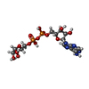
 Mass: 559.316 Da / Num. of mol.: 1 / Source method: obtained synthetically / Formula: C15H23N5O14P2 / Feature type: SUBJECT OF INVESTIGATION
 Mass: 18.015 Da / Num. of mol.: 211 / Source method: isolated from a natural source / Formula: H2O
 Sample preparation
 / Beamline: MASSIF-1 / Wavelength: 0.9655 Å
 Processing