 Movie
Movie Controller

[English] 日本語
 Yorodumi
- PDB-9qq4: Mini-bacterioferritin from Candidatus Methanoperedens species BLZ... -
+ Open data

- Basic information
| Entry | Database: PDB / ID: 9qq4 | |||||||||||||||||||||
|---|---|---|---|---|---|---|---|---|---|---|---|---|---|---|---|---|---|---|---|---|---|---|
| Title | Mini-bacterioferritin from Candidatus Methanoperedens species BLZ2 as isolated form at 1.07-A resolution | |||||||||||||||||||||
 Components Components | Bacterioferritin | |||||||||||||||||||||
 Keywords Keywords | OXIDOREDUCTASE / ferritin-like protein / mini-ferritin / Fe-coproporphyrin III / anaerobic methane-oxidising archaea / archaea / iron homeostasis / iron storage / dodecamer / nano-compartment | |||||||||||||||||||||
| Function / homology | : / Chem-FEC Function and homology information Function and homology information | |||||||||||||||||||||
| Biological species |  Candidatus Methanoperedens sp. BLZ2 (archaea) Candidatus Methanoperedens sp. BLZ2 (archaea) | |||||||||||||||||||||
| Method |  X-RAY DIFFRACTION / SYNCHROTRON / MOLECULAR REPLACEMENT / Resolution: 1.07 Å X-RAY DIFFRACTION / SYNCHROTRON / MOLECULAR REPLACEMENT / Resolution: 1.07 Å | |||||||||||||||||||||
 Authors Authors | Wissink, M. / Wagner, T. | |||||||||||||||||||||
| Funding support | European Union,  Germany, Germany,  Netherlands, 6items Netherlands, 6items
| |||||||||||||||||||||
 Citation Citation | Journal: Commun Biol / Year: 2026 Title: Mini-bacterioferritins: structural insight into a ferritin-like protein from the anaerobic methane-oxidising archaeon Candidatus Methanoperedens carboxydivorans. Authors: Wissink, M. / Engilberge, S. / Leao, P. / Jansen, R.S. / Jetten, M.S.M. / Belhamri, M. / Lemaire, O.N. / Royant, A. / Welte, C.U. / Wagner, T. | |||||||||||||||||||||
| History |
|
- Structure visualization
| Structure viewer | Molecule: MolmilJmol/JSmol |
|---|
- Downloads & links
-Download
| PDBx/mmCIF format | 9qq4.cif.gz | 402.7 KB | Display | PDBx/mmCIF format |
|---|---|---|---|---|
| PDB format | pdb9qq4.ent.gz | 338.2 KB | Display | PDB format |
| PDBx/mmJSON format | 9qq4.json.gz | Tree view | PDBx/mmJSON format | |
| Others |  Other downloads Other downloads |
-Validation report
| Arichive directory | https://data.pdbj.org/pub/pdb/validation_reports/qq/9qq4ftp://data.pdbj.org/pub/pdb/validation_reports/qq/9qq4 | HTTPS FTP |
|---|
-Related structure data


-Links
 PDBj
PDBj- Assembly
| Deposited unit | 
| ||||||||||||||||||
|---|---|---|---|---|---|---|---|---|---|---|---|---|---|---|---|---|---|---|---|
| 1 | 
| ||||||||||||||||||
| Unit cell |
| ||||||||||||||||||
| Components on special symmetry positions |
|
-Components
-Protein , 1 types, 4 molecules ABCD
| #1: Protein | Mass: 15969.113 Da / Num. of mol.: 4 / Source method: isolated from a natural source Details: Mini-bacterioferritin from Candidatus Methanoperedens species BLZ2 Source: (natural) Candidatus Methanoperedens sp. BLZ2 (archaea)Cell line: / / Organ: / / Plasmid details: / / Strain: BLZ2 / Tissue: / |
|---|
-Non-polymers , 7 types, 953 molecules 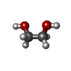




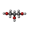

| #2: Chemical | ChemComp-EDO / 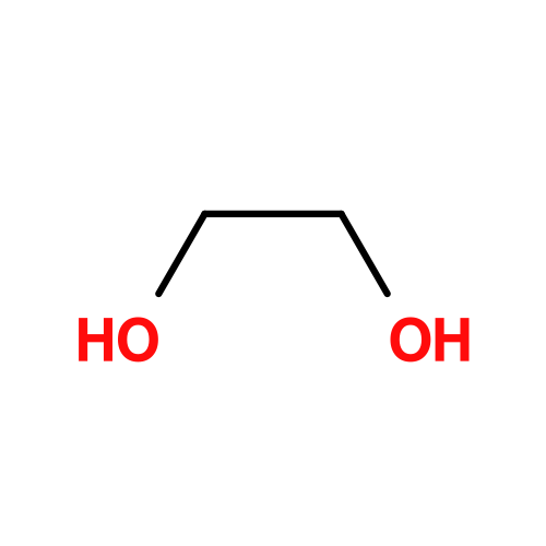 Mass: 62.068 Da / Num. of mol.: 8 / Source method: isolated from a natural source / Formula: C2H6O2 Mass: 62.068 Da / Num. of mol.: 8 / Source method: isolated from a natural source / Formula: C2H6O2#3: Chemical | ChemComp-FE /  Mass: 55.845 Da / Num. of mol.: 8 / Source method: obtained synthetically / Formula: Fe / Feature type: SUBJECT OF INVESTIGATION Mass: 55.845 Da / Num. of mol.: 8 / Source method: obtained synthetically / Formula: Fe / Feature type: SUBJECT OF INVESTIGATION#4: Chemical | ChemComp-FEC /  Mass: 708.538 Da / Num. of mol.: 4 / Source method: isolated from a natural source / Formula: C36H36FeN4O8 / Feature type: SUBJECT OF INVESTIGATION Mass: 708.538 Da / Num. of mol.: 4 / Source method: isolated from a natural source / Formula: C36H36FeN4O8 / Feature type: SUBJECT OF INVESTIGATION#5: Chemical | ChemComp-MG /  Mass: 24.305 Da / Num. of mol.: 4 / Source method: isolated from a natural source / Formula: Mg Mass: 24.305 Da / Num. of mol.: 4 / Source method: isolated from a natural source / Formula: Mg#6: Chemical | ChemComp-NA / 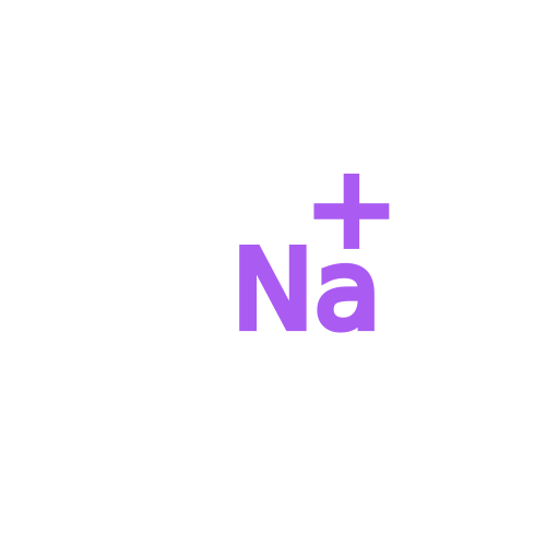 Mass: 22.990 Da / Num. of mol.: 4 / Source method: obtained synthetically / Formula: Na Mass: 22.990 Da / Num. of mol.: 4 / Source method: obtained synthetically / Formula: Na#7: Chemical | ChemComp-GOL / | 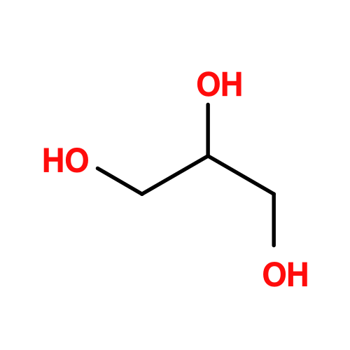 Mass: 92.094 Da / Num. of mol.: 1 / Source method: obtained synthetically / Formula: C3H8O3 Mass: 92.094 Da / Num. of mol.: 1 / Source method: obtained synthetically / Formula: C3H8O3#8: Water | ChemComp-HOH / | Mass: 18.015 Da / Num. of mol.: 924 / Source method: isolated from a natural source / Formula: H2O |
|---|
-Details
| Has ligand of interest | Y |
|---|---|
| Has protein modification | N |
-Experimental details
-Experiment
| Experiment | Method: X-RAY DIFFRACTION / Number of used crystals: 1 |
|---|
- Sample preparation
| Crystal | Density Matthews: 2.31 Å3/Da / Density % sol: 46.83 % / Description: Light pink bipyramids |
|---|---|
| Crystal grow | Temperature: 293.15 K / Method: vapor diffusion, sitting drop / pH: 7.5 Details: The crystal was obtained from an initial screening using the sitting drop method performed on a 96-well MRC 2-well polystyrene crystallisation plate (SWISSCI, Switzerland) at 20 degree ...Details: The crystal was obtained from an initial screening using the sitting drop method performed on a 96-well MRC 2-well polystyrene crystallisation plate (SWISSCI, Switzerland) at 20 degree Celsius under a pure N2 atmosphere. The plate was filled with 90 uL crystallisation solution in the large reservoir and the purified protein solution concentrated to 4.4 mg/mL was spotted in the small reservoirs by mixing 0.5 uL purified protein with 0.5 uL crystallisation solution using an OryxNano robot (Douglas Instruments Ltd, UK). The crystallisation solution was composed of: 20 % v/v Jeffamine M-600 pH 7.0, and 100 mM HEPES pH 7.5. Once prepared, the plate was transferred to an anaerobic chamber containing an N2/H2 (97/3 percent) atmosphere for long-term storage. Prior liquid nitrogen freezing, the crystal was soaked in the crystallization solution supplemented with 25 % v/v glycerol for a few seconds. PH range: 7.0-7.5 |
-Data collection
| Diffraction | Mean temperature: 100 K / Ambient temp details: / / Serial crystal experiment: N |
|---|---|
| Diffraction source | Source: SYNCHROTRON / Site: SLS  / Beamline: X06SA / Wavelength: 1 Å / Beamline: X06SA / Wavelength: 1 Å |
| Detector | Type: DECTRIS EIGER X 16M / Detector: PIXEL / Date: Dec 2, 2022 |
| Radiation | Protocol: SINGLE WAVELENGTH / Monochromatic (M) / Laue (L): M / Scattering type: x-ray |
| Radiation wavelength | Wavelength: 1 Å / Relative weight: 1 |
| Reflection | Resolution: 1.067→75.871 Å / Num. obs: 221484 / % possible obs: 95.8 % / Redundancy: 15.7 % / CC1/2: 0.999 / Rmerge(I) obs: 0.082 / Rpim(I) all: 0.02 / Rrim(I) all: 0.084 / Net I/σ(I): 18.5 |
| Reflection shell | Resolution: 1.067→1.129 Å / Redundancy: 5.9 % / Rmerge(I) obs: 1.052 / Mean I/σ(I) obs: 1.7 / Num. unique obs: 11074 / CC1/2: 0.654 / Rpim(I) all: 0.466 / Rrim(I) all: 1.154 / % possible all: 58.4 |
- Processing
| Software |
| |||||||||||||||||||||||||||||||||||||||||||||||||||||||||||||||||||||||||||||||||||||||||||||||||||||||||||||||||||||||||||||||||||||||||||||||||||||||||||||||||||||||||||||||||||||||||||||||||||||||||||||||||||||||||
|---|---|---|---|---|---|---|---|---|---|---|---|---|---|---|---|---|---|---|---|---|---|---|---|---|---|---|---|---|---|---|---|---|---|---|---|---|---|---|---|---|---|---|---|---|---|---|---|---|---|---|---|---|---|---|---|---|---|---|---|---|---|---|---|---|---|---|---|---|---|---|---|---|---|---|---|---|---|---|---|---|---|---|---|---|---|---|---|---|---|---|---|---|---|---|---|---|---|---|---|---|---|---|---|---|---|---|---|---|---|---|---|---|---|---|---|---|---|---|---|---|---|---|---|---|---|---|---|---|---|---|---|---|---|---|---|---|---|---|---|---|---|---|---|---|---|---|---|---|---|---|---|---|---|---|---|---|---|---|---|---|---|---|---|---|---|---|---|---|---|---|---|---|---|---|---|---|---|---|---|---|---|---|---|---|---|---|---|---|---|---|---|---|---|---|---|---|---|---|---|---|---|---|---|---|---|---|---|---|---|---|---|---|---|---|---|---|---|---|
| Refinement | Method to determine structure: MOLECULAR REPLACEMENT / Resolution: 1.07→75.87 Å / SU ML: 0.07 / Cross valid method: FREE R-VALUE / σ(F): 1.35 / Phase error: 10.64 / Stereochemistry target values: ML Details: The model was manually optimised with Coot (v0.9.8). Refinement was performed with Phenix refine (v1.21.1-5286) (Liebschner et al., 2019) without applying non-crystallography symmetry. The ...Details: The model was manually optimised with Coot (v0.9.8). Refinement was performed with Phenix refine (v1.21.1-5286) (Liebschner et al., 2019) without applying non-crystallography symmetry. The model was refined by considering all atoms anisotropic with hydrogens in riding positions. The model was validated by the molprobity tool integrated with PHENIX (Emsley et al., 2010; Liebschner et al., 2019) and with the oline MolProbity server (http://molprobity.biochem.duke.edu).
| |||||||||||||||||||||||||||||||||||||||||||||||||||||||||||||||||||||||||||||||||||||||||||||||||||||||||||||||||||||||||||||||||||||||||||||||||||||||||||||||||||||||||||||||||||||||||||||||||||||||||||||||||||||||||
| Solvent computation | Shrinkage radii: 0.9 Å / VDW probe radii: 1.1 Å / Solvent model: FLAT BULK SOLVENT MODEL | |||||||||||||||||||||||||||||||||||||||||||||||||||||||||||||||||||||||||||||||||||||||||||||||||||||||||||||||||||||||||||||||||||||||||||||||||||||||||||||||||||||||||||||||||||||||||||||||||||||||||||||||||||||||||
| Displacement parameters | Biso mean: 13.31 Å2 | |||||||||||||||||||||||||||||||||||||||||||||||||||||||||||||||||||||||||||||||||||||||||||||||||||||||||||||||||||||||||||||||||||||||||||||||||||||||||||||||||||||||||||||||||||||||||||||||||||||||||||||||||||||||||
| Refinement step | Cycle: LAST / Resolution: 1.07→75.87 Å
| |||||||||||||||||||||||||||||||||||||||||||||||||||||||||||||||||||||||||||||||||||||||||||||||||||||||||||||||||||||||||||||||||||||||||||||||||||||||||||||||||||||||||||||||||||||||||||||||||||||||||||||||||||||||||
| Refine LS restraints |
| |||||||||||||||||||||||||||||||||||||||||||||||||||||||||||||||||||||||||||||||||||||||||||||||||||||||||||||||||||||||||||||||||||||||||||||||||||||||||||||||||||||||||||||||||||||||||||||||||||||||||||||||||||||||||
| LS refinement shell |
|
