 Movie
Movie Controller

[English] 日本語
 Yorodumi
- PDB-9qlu: Amyloid structure of 17kDa alpha-amylase/trypsin inhibitor 2 (Uni... -
+ Open data

- Basic information
| Entry | Database: PDB / ID: 9qlu | |||||||||||||||||||||||||||
|---|---|---|---|---|---|---|---|---|---|---|---|---|---|---|---|---|---|---|---|---|---|---|---|---|---|---|---|---|
| Title | Amyloid structure of 17kDa alpha-amylase/trypsin inhibitor 2 (Uniprot ID: AI172_ORYSJ) | |||||||||||||||||||||||||||
 Components Components | 17kDa alpha-amylase/trypsin inhibitor 2 | |||||||||||||||||||||||||||
 Keywords Keywords | PROTEIN FIBRIL / Amyloid / Plant protein / Storage protein / Trypsin inhibitor | |||||||||||||||||||||||||||
| Function / homology | Cereal seed allergen/grain softness/trypsin and alpha-amylase inhibitor / Protease inhibitor/seed storage/LTP family / Bifunctional inhibitor/plant lipid transfer protein/seed storage helical domain / Bifunctional inhibitor/plant lipid transfer protein/seed storage helical domain superfamily / nutrient reservoir activity / IgE binding / serine-type endopeptidase inhibitor activity / extracellular region / 17kDa alpha-amylase/trypsin inhibitor 2 Function and homology information Function and homology information | |||||||||||||||||||||||||||
| Biological species |  | |||||||||||||||||||||||||||
| Method | ELECTRON MICROSCOPY / helical reconstruction / cryo EM / Resolution: 2.54 Å | |||||||||||||||||||||||||||
 Authors Authors | Rhyner, D. / Riek, R. / Greenwald, J. / Frey, L. / Kwiatkowski, W. | |||||||||||||||||||||||||||
| Funding support |  Switzerland, 1items Switzerland, 1items
| |||||||||||||||||||||||||||
 Citation Citation | Journal: Protein Sci / Year: 2025 Title: Impurities in amyloid studies: The power of automated model building within a cautionary tale for structural biologists. Authors: David Rhyner / Lukas Frey / Jiangtao Zhou / Witek Kwiatkowski / Raffaele Mezzenga / Roland Riek / Jason Greenwald / Abstract: The purity of protein samples of biological origin is often difficult to ascertain, leading the naïve or optimistic scientist to underestimate contaminants in their research. Even after extensive ...The purity of protein samples of biological origin is often difficult to ascertain, leading the naïve or optimistic scientist to underestimate contaminants in their research. Even after extensive purification, protein samples can contain nucleic acids, truncated degradation products, or other protein contaminants. While in many cases, and when present at low concentrations, such contaminants are unlikely to alter experimental results significantly, they must be considered when studying protein aggregation. Such reactions can be sensitive to small environmental changes in their early stages due to a nucleation-dependent mechanism, where minor differences can be amplified during the subsequent exponential growth phase. During a recent study of the amyloid formation of human lysozyme, we encountered a significant amyloid-forming protein contaminant derived from the expression host Oryza sativa japonica. Further investigation of this widely used commercial source of human lysozyme revealed at least a dozen protein contaminants. These discoveries led to intriguing observations, including an underdeveloped branch of plant amyloid research and a possible link between the amyloid fold and allergens. Here, we present our findings within a cautionary tale for structural biologists: a surprising variety of contaminants in a commercial protein sample and the accidental yet definitive identification of one of them by cryo-electron microscopy helical reconstruction. The resulting 2.54 Å model of the 17 kDa alpha-amylase/trypsin inhibitor Type 2 marks the first known amyloid structure of a plant protein. | |||||||||||||||||||||||||||
| History |
|
- Structure visualization
| Structure viewer | Molecule: MolmilJmol/JSmol |
|---|
- Downloads & links
-Download
| PDBx/mmCIF format | 9qlu.cif.gz | 73.8 KB | Display | PDBx/mmCIF format |
|---|---|---|---|---|
| PDB format | pdb9qlu.ent.gz | 52.2 KB | Display | PDB format |
| PDBx/mmJSON format | 9qlu.json.gz | Tree view | PDBx/mmJSON format | |
| Others |  Other downloads Other downloads |
-Validation report
| Arichive directory | https://data.pdbj.org/pub/pdb/validation_reports/ql/9qluftp://data.pdbj.org/pub/pdb/validation_reports/ql/9qlu | HTTPS FTP |
|---|
-Related structure data
| Related structure data | 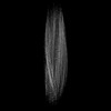 53234MC M: map data used to model this data C: citing same article ( |
|---|---|
| Similar structure data |
-Links
 PDBj
PDBj

- Assembly
| Deposited unit | 
|
|---|---|
| 1 |
|
-Components
| #1: Protein | Mass: 16493.027 Da / Num. of mol.: 5 Source method: isolated from a genetically manipulated source Source: (gene. exp.) Gene: Os07g0216700, LOC_Os07g11650, OJ1080_F08.106, OJ1779_B07.133, OsJ_23556 Production host: References: UniProt: Q7X8H9 Has protein modification | N | |
|---|
-Experimental details
-Experiment
| Experiment | Method: ELECTRON MICROSCOPY |
|---|---|
| EM experiment | Aggregation state: FILAMENT / 3D reconstruction method: helical reconstruction |
- Sample preparation
| Component | Name: Amyloid fibril of 17kDa alpha-amylase/trypsin inhibitor 2 Type: ORGANELLE OR CELLULAR COMPONENT / Entity ID: all / Source: NATURAL |
|---|---|
| Molecular weight | Experimental value: NO |
| Source (natural) | Organism: |
| Buffer solution | pH: 7 / Details: DTT 100 mM |
| Specimen | Embedding applied: NO / Shadowing applied: NO / Staining applied: NO / Vitrification applied: YES |
| Specimen support | Grid material: COPPER / Grid type: Quantifoil R2/1 |
| Vitrification | Instrument: FEI VITROBOT MARK IV / Cryogen name: ETHANE-PROPANE / Humidity: 95 % / Chamber temperature: 288.15 K |
- Electron microscopy imaging
| Experimental equipment |  Model: Titan Krios / Image courtesy: FEI Company |
|---|---|
| Microscopy | Model: TFS KRIOS |
| Electron gun | Electron source:  FIELD EMISSION GUN / Accelerating voltage: 300 kV / Illumination mode: FLOOD BEAM FIELD EMISSION GUN / Accelerating voltage: 300 kV / Illumination mode: FLOOD BEAM |
| Electron lens | Mode: BRIGHT FIELD / Nominal defocus max: 3000 nm / Nominal defocus min: 800 nm / Cs: 2.7 mm / C2 aperture diameter: 70 µm / Alignment procedure: COMA FREE |
| Specimen holder | Cryogen: NITROGEN / Specimen holder model: FEI TITAN KRIOS AUTOGRID HOLDER |
| Image recording | Average exposure time: 1 sec. / Electron dose: 62.79 e/Å2 / Film or detector model: GATAN K3 BIOQUANTUM (6k x 4k) |
| EM imaging optics | Energyfilter name: GIF Bioquantum / Energyfilter slit width: 20 eV |
- Processing
| EM software | Name: RELION / Version: 4.0.0 / Category: 3D reconstruction |
|---|---|
| CTF correction | Type: PHASE FLIPPING AND AMPLITUDE CORRECTION |
| Helical symmerty | Angular rotation/subunit: -2.277 ° / Axial rise/subunit: 4.734 Å / Axial symmetry: C1 |
| 3D reconstruction | Resolution: 2.54 Å / Resolution method: FSC 0.143 CUT-OFF / Num. of particles: 121923 / Symmetry type: HELICAL |
