 Movie
Movie Controller


+ Open data

- Basic information
| Entry | Database: PDB / ID: 9qfp | |||||||||||||||
|---|---|---|---|---|---|---|---|---|---|---|---|---|---|---|---|---|
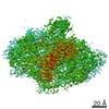
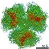
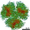
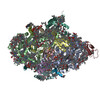
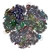
| Title | Monomeric Photosystem I Cryo-EM structure at 1.8 A resolution | |||||||||||||||
 Components Components | (Photosystem I ...) x 11 | |||||||||||||||
 Keywords Keywords | PHOTOSYNTHESIS / Monomeric Photosystem I / mPSI | |||||||||||||||
| Function / homology |  Function and homology information Function and homology informationphotosystem I reaction center / photosystem I / photosynthetic electron transport in photosystem I / photosystem I / plasma membrane-derived thylakoid membrane / photosynthesis / 4 iron, 4 sulfur cluster binding / electron transfer activity / magnesium ion binding Similarity search - Function | |||||||||||||||
| Biological species |   Thermosynechococcus vestitus BP-1 (bacteria) Thermosynechococcus vestitus BP-1 (bacteria) | |||||||||||||||
| Method | ELECTRON MICROSCOPY / single particle reconstruction / cryo EM / Resolution: 1.74 Å | |||||||||||||||
 Authors Authors | Gaullier, G. / Boyka, J. / Zouni, A. / Blikstad, C. | |||||||||||||||
| Funding support |  Germany, Germany,  Sweden, 4items Sweden, 4items
| |||||||||||||||
 Citation Citation | Journal: To Be Published Title: Monomeric Photosystem I Cryo-EM structure at 1.8 A resolution Authors: Boyka, J. / Gaullier, G. / Blikstad, C. / Zouni, A. | |||||||||||||||
| History |
|
- Structure visualization
| Structure viewer | Molecule: MolmilJmol/JSmol |
|---|
- Downloads & links
-Download
| PDBx/mmCIF format | 9qfp.cif.gz | 653.1 KB | Display | PDBx/mmCIF format |
|---|---|---|---|---|
| PDB format | pdb9qfp.ent.gz | 567.2 KB | Display | PDB format |
| PDBx/mmJSON format | 9qfp.json.gz | Tree view | PDBx/mmJSON format | |
| Others |  Other downloads Other downloads |
-Validation report
| Arichive directory | https://data.pdbj.org/pub/pdb/validation_reports/qf/9qfpftp://data.pdbj.org/pub/pdb/validation_reports/qf/9qfp | HTTPS FTP |
|---|
-Related structure data
| Related structure data |  53120MC M: map data used to model this data C: citing same article ( |
|---|---|
| Similar structure data |

-Links
 PDBj
PDBj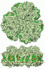

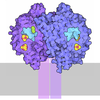
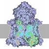
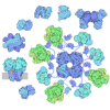

- Assembly
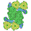
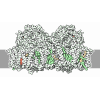
| Deposited unit | 
|
|---|---|
| 1 |
|
-Components
-Photosystem I ... , 11 types, 11 molecules ABCDEFIJKMX
| #1: Protein | Mass: 83267.773 Da / Num. of mol.: 1 / Source method: isolated from a natural source Source: (natural) Thermosynechococcus vestitus BP-1 (bacteria)References: UniProt: P0A405, photosystem I |
|---|---|
| #2: Protein | Mass: 83123.648 Da / Num. of mol.: 1 / Source method: isolated from a natural source Source: (natural) Thermosynechococcus vestitus BP-1 (bacteria)References: UniProt: P0A407, photosystem I |
| #3: Protein | Mass: 8809.207 Da / Num. of mol.: 1 / Source method: isolated from a natural source Source: (natural) Thermosynechococcus vestitus BP-1 (bacteria)References: UniProt: P0A415, photosystem I |
| #4: Protein | Mass: 15389.494 Da / Num. of mol.: 1 / Source method: isolated from a natural source Source: (natural) Thermosynechococcus vestitus BP-1 (bacteria)References: UniProt: P0A420 |
| #5: Protein | Mass: 8399.485 Da / Num. of mol.: 1 / Source method: isolated from a natural source Source: (natural) Thermosynechococcus vestitus BP-1 (bacteria)References: UniProt: P0A423 |
| #6: Protein | Mass: 17716.586 Da / Num. of mol.: 1 / Source method: isolated from a natural source Source: (natural) Thermosynechococcus vestitus BP-1 (bacteria)References: UniProt: P0A401 |
| #7: Protein/peptide | Mass: 4297.234 Da / Num. of mol.: 1 / Source method: isolated from a natural source Source: (natural) Thermosynechococcus vestitus BP-1 (bacteria)References: UniProt: P0A427 |
| #8: Protein/peptide | Mass: 4770.698 Da / Num. of mol.: 1 / Source method: isolated from a natural source Source: (natural) Thermosynechococcus vestitus BP-1 (bacteria)References: UniProt: P0A429 |
| #9: Protein | Mass: 8483.983 Da / Num. of mol.: 1 / Source method: isolated from a natural source Source: (natural) Thermosynechococcus vestitus BP-1 (bacteria)References: UniProt: P0A425 |
| #10: Protein/peptide | Mass: 3426.115 Da / Num. of mol.: 1 / Source method: isolated from a natural source Source: (natural) Thermosynechococcus vestitus BP-1 (bacteria)References: UniProt: P0A403 |
| #11: Protein/peptide | Mass: 4424.317 Da / Num. of mol.: 1 / Source method: isolated from a natural source Source: (natural) Thermosynechococcus vestitus BP-1 (bacteria)References: UniProt: Q8DKP6 |
-Sugars , 1 types, 16 molecules 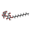
| #12: Sugar | ChemComp-LMT / 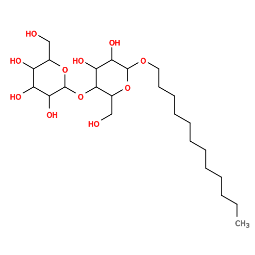 Type: D-saccharide / Mass: 510.615 Da / Num. of mol.: 16 / Source method: obtained synthetically / Formula: C24H46O11 / Feature type: SUBJECT OF INVESTIGATION / Comment: detergent*YM Type: D-saccharide / Mass: 510.615 Da / Num. of mol.: 16 / Source method: obtained synthetically / Formula: C24H46O11 / Feature type: SUBJECT OF INVESTIGATION / Comment: detergent*YM |
|---|
-Non-polymers , 10 types, 1306 molecules 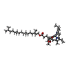
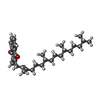
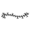
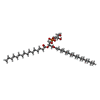
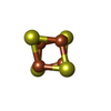


| #13: Chemical | ChemComp-CLA / 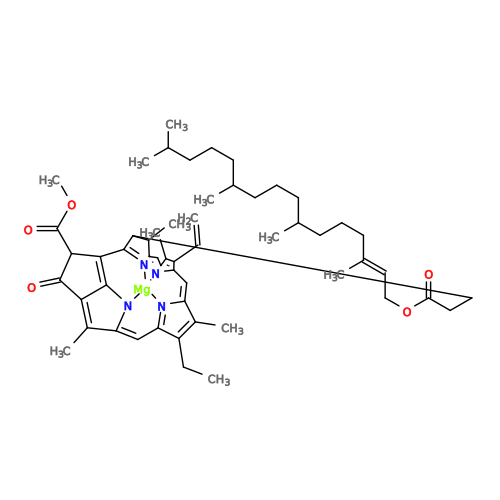 Mass: 893.489 Da / Num. of mol.: 90 / Source method: obtained synthetically / Formula: C55H72MgN4O5 / Feature type: SUBJECT OF INVESTIGATION Mass: 893.489 Da / Num. of mol.: 90 / Source method: obtained synthetically / Formula: C55H72MgN4O5 / Feature type: SUBJECT OF INVESTIGATION#14: Chemical | 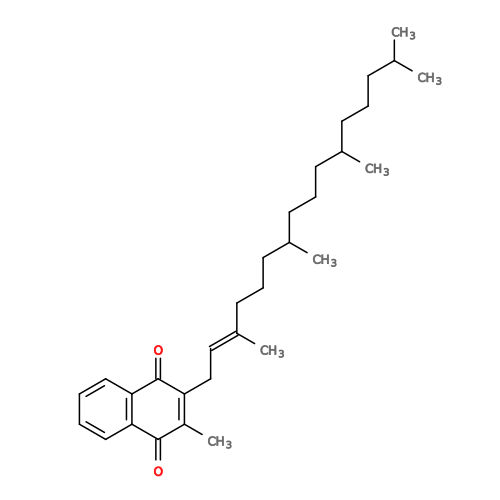 Mass: 450.696 Da / Num. of mol.: 2 / Source method: obtained synthetically / Formula: C31H46O2 / Feature type: SUBJECT OF INVESTIGATION Mass: 450.696 Da / Num. of mol.: 2 / Source method: obtained synthetically / Formula: C31H46O2 / Feature type: SUBJECT OF INVESTIGATION#15: Chemical | ChemComp-BCR / 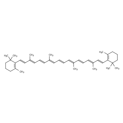 Mass: 536.873 Da / Num. of mol.: 19 / Source method: obtained synthetically / Formula: C40H56 / Feature type: SUBJECT OF INVESTIGATION Mass: 536.873 Da / Num. of mol.: 19 / Source method: obtained synthetically / Formula: C40H56 / Feature type: SUBJECT OF INVESTIGATION#16: Chemical | ChemComp-LHG / 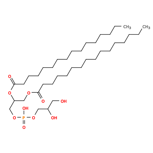 Mass: 722.970 Da / Num. of mol.: 5 / Source method: obtained synthetically / Formula: C38H75O10P / Feature type: SUBJECT OF INVESTIGATION / Comment: phospholipid*YM Mass: 722.970 Da / Num. of mol.: 5 / Source method: obtained synthetically / Formula: C38H75O10P / Feature type: SUBJECT OF INVESTIGATION / Comment: phospholipid*YM#17: Chemical | 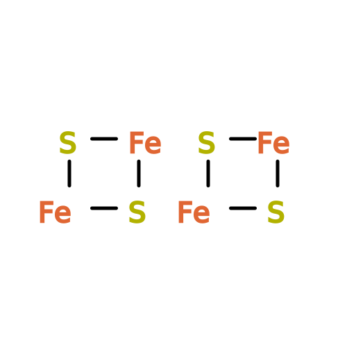 Mass: 351.640 Da / Num. of mol.: 3 / Source method: obtained synthetically / Formula: Fe4S4 / Feature type: SUBJECT OF INVESTIGATION Mass: 351.640 Da / Num. of mol.: 3 / Source method: obtained synthetically / Formula: Fe4S4 / Feature type: SUBJECT OF INVESTIGATION#18: Chemical | ChemComp-CL0 / | 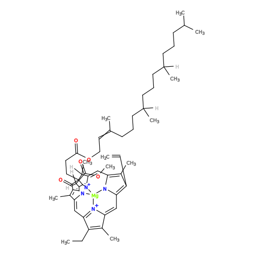 Mass: 893.489 Da / Num. of mol.: 1 / Source method: obtained synthetically / Formula: C55H72MgN4O5 Mass: 893.489 Da / Num. of mol.: 1 / Source method: obtained synthetically / Formula: C55H72MgN4O5#19: Chemical | ChemComp-LMG / | 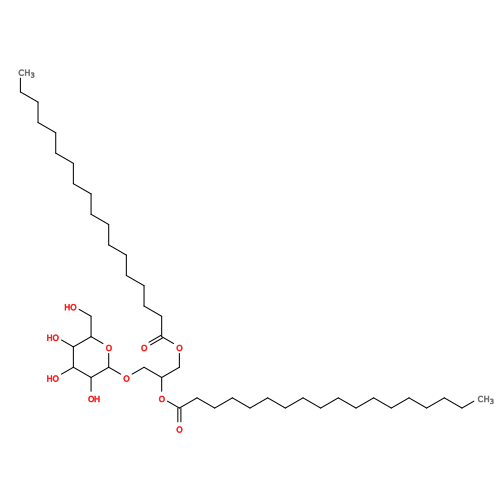 Mass: 787.158 Da / Num. of mol.: 1 / Source method: obtained synthetically / Formula: C45H86O10 / Feature type: SUBJECT OF INVESTIGATION Mass: 787.158 Da / Num. of mol.: 1 / Source method: obtained synthetically / Formula: C45H86O10 / Feature type: SUBJECT OF INVESTIGATION#20: Chemical | ChemComp-MG / |  Mass: 24.305 Da / Num. of mol.: 1 / Source method: obtained synthetically / Formula: Mg / Feature type: SUBJECT OF INVESTIGATION Mass: 24.305 Da / Num. of mol.: 1 / Source method: obtained synthetically / Formula: Mg / Feature type: SUBJECT OF INVESTIGATION#21: Chemical | ChemComp-SQD / | 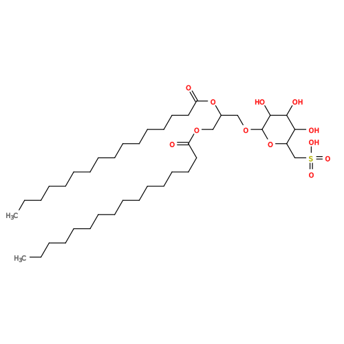 Mass: 795.116 Da / Num. of mol.: 1 / Source method: obtained synthetically / Formula: C41H78O12S / Feature type: SUBJECT OF INVESTIGATION Mass: 795.116 Da / Num. of mol.: 1 / Source method: obtained synthetically / Formula: C41H78O12S / Feature type: SUBJECT OF INVESTIGATION#22: Water | ChemComp-HOH / | Mass: 18.015 Da / Num. of mol.: 1183 / Source method: isolated from a natural source / Formula: H2O |
|---|
-Details
| Has ligand of interest | Y |
|---|---|
| Has protein modification | Y |
-Experimental details
-Experiment
| Experiment | Method: ELECTRON MICROSCOPY |
|---|---|
| EM experiment | Aggregation state: PARTICLE / 3D reconstruction method: single particle reconstruction |
- Sample preparation
| Component | Name: Monomeric photosystem I / Type: COMPLEX Details: Endogeneous photosystem I complex purified from its native source organism Thermosynechococcus vestitus BP-1. Entity ID: #1-#11 / Source: NATURAL | ||||||||||||||||||||
|---|---|---|---|---|---|---|---|---|---|---|---|---|---|---|---|---|---|---|---|---|---|
| Molecular weight | Experimental value: NO | ||||||||||||||||||||
| Source (natural) | Organism: Thermosynechococcus vestitus BP-1 (bacteria) | ||||||||||||||||||||
| Buffer solution | pH: 8 | ||||||||||||||||||||
| Buffer component |
| ||||||||||||||||||||
| Specimen | Conc.: 2.5 mg/ml / Embedding applied: NO / Shadowing applied: NO / Staining applied: NO / Vitrification applied: YES / Details: Concentration is expressed in mM chlorophyll. | ||||||||||||||||||||
| Specimen support | Details: Current 20 mA. Performed with a Pelco easiGlow device. Grid material: COPPER / Grid mesh size: 300 divisions/in. / Grid type: Quantifoil R1.2/1.3 | ||||||||||||||||||||
| Vitrification | Instrument: FEI VITROBOT MARK IV / Cryogen name: ETHANE / Humidity: 100 % / Chamber temperature: 277 K Details: Delay time 30 s Blot time 2 s Blot force 0 Drain time 0 s |
- Electron microscopy imaging
| Experimental equipment |  Model: Titan Krios / Image courtesy: FEI Company |
|---|---|
| Microscopy | Model: TFS KRIOS |
| Electron gun | Electron source:  FIELD EMISSION GUN / Accelerating voltage: 300 kV / Illumination mode: FLOOD BEAM FIELD EMISSION GUN / Accelerating voltage: 300 kV / Illumination mode: FLOOD BEAM |
| Electron lens | Mode: BRIGHT FIELD / Nominal magnification: 130000 X / Nominal defocus max: 1400 nm / Nominal defocus min: 400 nm / Cs: 2.7 mm / Alignment procedure: COMA FREE |
| Specimen holder | Cryogen: NITROGEN / Specimen holder model: FEI TITAN KRIOS AUTOGRID HOLDER |
| Image recording | Electron dose: 40 e/Å2 / Film or detector model: GATAN K3 BIOQUANTUM (6k x 4k) / Num. of grids imaged: 1 / Num. of real images: 22001 |
| EM imaging optics | Energyfilter name: GIF Bioquantum / Energyfilter slit width: 20 eV |
- Processing
| EM software |
| ||||||||||||||||||||||||||||||||||||||||||||||||||||||||||||||||||||||||||||||||||||
|---|---|---|---|---|---|---|---|---|---|---|---|---|---|---|---|---|---|---|---|---|---|---|---|---|---|---|---|---|---|---|---|---|---|---|---|---|---|---|---|---|---|---|---|---|---|---|---|---|---|---|---|---|---|---|---|---|---|---|---|---|---|---|---|---|---|---|---|---|---|---|---|---|---|---|---|---|---|---|---|---|---|---|---|---|---|
| CTF correction | Details: Patch CTF estimation job in CryoSPARC, run with default settings. Type: PHASE FLIPPING AND AMPLITUDE CORRECTION | ||||||||||||||||||||||||||||||||||||||||||||||||||||||||||||||||||||||||||||||||||||
| Particle selection | Num. of particles selected: 1000224 Details: Topaz was trained with a manually picked set of 1010 particles. | ||||||||||||||||||||||||||||||||||||||||||||||||||||||||||||||||||||||||||||||||||||
| Symmetry | Point symmetry: C1 (asymmetric) | ||||||||||||||||||||||||||||||||||||||||||||||||||||||||||||||||||||||||||||||||||||
| 3D reconstruction | Resolution: 1.74 Å / Resolution method: FSC 0.143 CUT-OFF / Num. of particles: 712866 / Algorithm: FOURIER SPACE Details: Resolution as reported by CryoSPARC, using its mask auto-tightening procedure. Symmetry type: POINT | ||||||||||||||||||||||||||||||||||||||||||||||||||||||||||||||||||||||||||||||||||||
| Atomic model building | Protocol: FLEXIBLE FIT / Space: REAL Details: Alphafold2 predictions fetched from AlphaFold-DB were placed into the map by rigid body fitting. Termini not supported by the map were trimmed. Every residue was visually inspected at least ...Details: Alphafold2 predictions fetched from AlphaFold-DB were placed into the map by rigid body fitting. Termini not supported by the map were trimmed. Every residue was visually inspected at least once, and its fit to density adjusted if necessary, using ISOLDE. | ||||||||||||||||||||||||||||||||||||||||||||||||||||||||||||||||||||||||||||||||||||
| Atomic model building |
|
