+ Open data
- Basic information
| Entry | Database: PDB / ID: 9qdq | ||||||||||||||||||
|---|---|---|---|---|---|---|---|---|---|---|---|---|---|---|---|---|---|---|---|
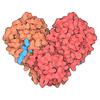
| Title | Cryo-EM structure of Upf1-Nmd4-Ebs1 in complex with RNA | ||||||||||||||||||
 Components Components |
| ||||||||||||||||||
 Keywords Keywords | RNA BINDING PROTEIN / helicase / Nonsense-mediated decay | ||||||||||||||||||
| Function / homology |  Function and homology information Function and homology informationcytoplasmic RNA surveillance / double-stranded DNA helicase activity / nuclear-transcribed mRNA catabolic process, 3'-5' exonucleolytic nonsense-mediated decay / intracellular mRNA localization / regulation of translational termination / telomerase holoenzyme complex / telomerase RNA binding / silent mating-type cassette heterochromatin formation / nuclear-transcribed mRNA catabolic process, nonsense-mediated decay / telomeric DNA binding ...cytoplasmic RNA surveillance / double-stranded DNA helicase activity / nuclear-transcribed mRNA catabolic process, 3'-5' exonucleolytic nonsense-mediated decay / intracellular mRNA localization / regulation of translational termination / telomerase holoenzyme complex / telomerase RNA binding / silent mating-type cassette heterochromatin formation / nuclear-transcribed mRNA catabolic process, nonsense-mediated decay / telomeric DNA binding / Nonsense Mediated Decay (NMD) independent of the Exon Junction Complex (EJC) / Nonsense Mediated Decay (NMD) enhanced by the Exon Junction Complex (EJC) / ribosomal small subunit binding / nuclear-transcribed mRNA catabolic process / P-body / single-stranded DNA binding / DNA recombination / DNA helicase / chromosome, telomeric region / RNA helicase activity / negative regulation of translation / protein ubiquitination / RNA helicase / mRNA binding / ATP hydrolysis activity / RNA binding / zinc ion binding / ATP binding / nucleus / cytoplasm Similarity search - Function | ||||||||||||||||||
| Biological species |  | ||||||||||||||||||
| Method | ELECTRON MICROSCOPY / single particle reconstruction / cryo EM / Resolution: 3.6 Å | ||||||||||||||||||
 Authors Authors | Iermak, I. / Wilson Eisele, N.R. / Kurscheidt, K. / Loukeri, M.J. / Basquin, J. / Bonneau, F. / Langer, L.M. / Keidel, A. / Conti, E. | ||||||||||||||||||
| Funding support |  Germany, European Union, Germany, European Union,  Denmark, 5items Denmark, 5items
| ||||||||||||||||||
 Citation Citation |  Journal: Cell Rep / Year: 2025 Journal: Cell Rep / Year: 2025Title: Molecular mechanisms governing the formation of distinct Upf1-containing complexes in yeast. Authors: Iuliia Iermak / Nicole R Wilson Eisele / Katharina Kurscheidt / Matina-Jasemi Loukeri / Jérôme Basquin / Fabien Bonneau / Lukas M Langer / Achim Keidel / Elena Conti /  Abstract: Upf1 is a master regulator of nonsense-mediated mRNA decay (NMD), an mRNA surveillance and degradation pathway conserved from yeast to human. In Saccharomyces cerevisiae, Upf1 exists in two distinct ...Upf1 is a master regulator of nonsense-mediated mRNA decay (NMD), an mRNA surveillance and degradation pathway conserved from yeast to human. In Saccharomyces cerevisiae, Upf1 exists in two distinct complexes with factors that mediate NMD activation or 5'-3' mRNA degradation. We combined endogenous purifications and biochemical reconstitutions of yeast Upf1 complexes with structural analyses and biochemical assays to elucidate the molecular mechanisms driving the organization of the Upf1-5'-3' and Upf1-2-3 complexes. We show that yeast Upf1 is in a constitutive complex, whereby its CH, RecA, and C-terminal domains interact with the mRNA decapping factor Dcp2, NMD-associated proteins Nmd4 and Ebs1, and the 5'-3' exoribonuclease Xrn1, respectively. Together, the interacting surfaces and closed conformation of Upf1 in the Upf1-5'-3' complex sterically obstruct the binding of Upf2-3. Our work points to a major restructuring upon recruitment of these factors during NMD and provides insights into evolutionary divergence amongst species. | ||||||||||||||||||
| History |
|
- Structure visualization
| Structure viewer | Molecule:  Molmil Molmil Jmol/JSmol Jmol/JSmol |
|---|
- Downloads & links
- Download
| PDBx/mmCIF format |  9qdq.cif.gz 9qdq.cif.gz | 274.8 KB | Display |  PDBx/mmCIF format PDBx/mmCIF format |
|---|---|---|---|---|
| PDB format |  pdb9qdq.ent.gz pdb9qdq.ent.gz | 211.1 KB | Display |  PDB format PDB format |
| PDBx/mmJSON format |  9qdq.json.gz 9qdq.json.gz | Tree view |  PDBx/mmJSON format PDBx/mmJSON format | |
| Others |  Other downloads Other downloads |
-Validation report
| Arichive directory |  https://data.pdbj.org/pub/pdb/validation_reports/qd/9qdq https://data.pdbj.org/pub/pdb/validation_reports/qd/9qdq ftp://data.pdbj.org/pub/pdb/validation_reports/qd/9qdq ftp://data.pdbj.org/pub/pdb/validation_reports/qd/9qdq | HTTPS FTP |
|---|
-Related structure data
| Related structure data |  53032MC M: map data used to model this data C: citing same article ( |
|---|---|
| Similar structure data | Similarity search - Function & homology  F&H Search F&H Search |
- Links
Links
- Assembly
| Deposited unit | 
|
|---|---|
| 1 |
|
- Components
| #1: Protein | Mass: 109561.008 Da / Num. of mol.: 1 Source method: isolated from a genetically manipulated source Source: (gene. exp.)  Gene: NAM7, IFS2, MOF4, UPF1, YMR080C, YM9582.05C / Production host:  Trichoplusia ni (cabbage looper) / References: UniProt: P30771, DNA helicase, RNA helicase Trichoplusia ni (cabbage looper) / References: UniProt: P30771, DNA helicase, RNA helicase |
|---|---|
| #2: Protein | Mass: 100102.453 Da / Num. of mol.: 1 Source method: isolated from a genetically manipulated source Source: (gene. exp.)  Gene: EBS1, YDR206W, YD8142.03 / Production host:  |
| #3: Protein | Mass: 25316.912 Da / Num. of mol.: 1 Source method: isolated from a genetically manipulated source Source: (gene. exp.)  Gene: NMD4, YLR363C / Production host:  |
| #4: RNA chain | Mass: 9140.017 Da / Num. of mol.: 1 / Source method: obtained synthetically / Source: (synth.)  |
| Has protein modification | N |
-Experimental details
-Experiment
| Experiment | Method: ELECTRON MICROSCOPY |
|---|---|
| EM experiment | Aggregation state: PARTICLE / 3D reconstruction method: single particle reconstruction |
- Sample preparation
| Component | Name: Complex of Upf1, Nmd4 and Ebs1 bound to RNA / Type: COMPLEX / Entity ID: all / Source: RECOMBINANT |
|---|---|
| Molecular weight | Experimental value: NO |
| Source (natural) | Organism:  |
| Source (recombinant) | Organism:  Trichoplusia ni (cabbage looper) Trichoplusia ni (cabbage looper) |
| Buffer solution | pH: 7.5 |
| Specimen | Embedding applied: NO / Shadowing applied: NO / Staining applied: NO / Vitrification applied: YES |
| Specimen support | Grid material: COPPER / Grid mesh size: 200 divisions/in. / Grid type: Quantifoil R2/1 |
| Vitrification | Cryogen name: ETHANE-PROPANE |
- Electron microscopy imaging
| Experimental equipment |  Model: Titan Krios / Image courtesy: FEI Company |
|---|---|
| Microscopy | Model: TFS KRIOS |
| Electron gun | Electron source:  FIELD EMISSION GUN / Accelerating voltage: 300 kV / Illumination mode: FLOOD BEAM FIELD EMISSION GUN / Accelerating voltage: 300 kV / Illumination mode: FLOOD BEAM |
| Electron lens | Mode: BRIGHT FIELD / Nominal defocus max: 2000 nm / Nominal defocus min: 500 nm |
| Image recording | Electron dose: 60 e/Å2 / Film or detector model: GATAN K3 BIOQUANTUM (6k x 4k) |
| EM imaging optics | Energyfilter name: GIF Bioquantum |
- Processing
| EM software |
| ||||||||||||||||||||||||
|---|---|---|---|---|---|---|---|---|---|---|---|---|---|---|---|---|---|---|---|---|---|---|---|---|---|
| CTF correction | Type: PHASE FLIPPING AND AMPLITUDE CORRECTION | ||||||||||||||||||||||||
| 3D reconstruction | Resolution: 3.6 Å / Resolution method: FSC 0.143 CUT-OFF / Num. of particles: 160049 / Symmetry type: POINT | ||||||||||||||||||||||||
| Atomic model building | Protocol: RIGID BODY FIT / Space: REAL Details: Initial fitting of the Alphafold3 model was done in ChimeraX. Then model was rigid body refined and real space refined in Phenix | ||||||||||||||||||||||||
| Atomic model building | Source name: AlphaFold / Type: in silico model | ||||||||||||||||||||||||
| Refinement | Highest resolution: 3.6 Å Stereochemistry target values: REAL-SPACE (WEIGHTED MAP SUM AT ATOM CENTERS) | ||||||||||||||||||||||||
| Refine LS restraints |
|
 Movie
Movie Controller


 PDBj
PDBj


