 Movie
Movie Controller

[English] 日本語
 Yorodumi
- PDB-9q7m: Barbed end of cofilin actin, cofilin on second-to-last barbed end... -
+ Open data

- Basic information
| Entry | Database: PDB / ID: 9q7m | ||||||||||||||||||||||||
|---|---|---|---|---|---|---|---|---|---|---|---|---|---|---|---|---|---|---|---|---|---|---|---|---|---|
| Title | Barbed end of cofilin actin, cofilin on second-to-last barbed end subunit | ||||||||||||||||||||||||
 Components Components |
| ||||||||||||||||||||||||
 Keywords Keywords | PROTEIN FIBRIL / Cytoskeleton / Actin / Filament / Helical | ||||||||||||||||||||||||
| Function / homology |  Function and homology information Function and homology informationactin filament fragmentation / positive regulation of actin filament depolymerization / actin filament severing / actin filament depolymerization / skeletal muscle tissue development / I band / cytoskeletal motor activator activity / myosin heavy chain binding / tropomyosin binding / actin filament bundle ...actin filament fragmentation / positive regulation of actin filament depolymerization / actin filament severing / actin filament depolymerization / skeletal muscle tissue development / I band / cytoskeletal motor activator activity / myosin heavy chain binding / tropomyosin binding / actin filament bundle / troponin I binding / filamentous actin / mesenchyme migration / skeletal muscle myofibril / striated muscle thin filament / actin filament bundle assembly / skeletal muscle thin filament assembly / actin monomer binding / skeletal muscle fiber development / actin filament polymerization / stress fiber / titin binding / filopodium / actin filament / Hydrolases; Acting on acid anhydrides; Acting on acid anhydrides to facilitate cellular and subcellular movement / Z disc / nuclear matrix / calcium-dependent protein binding / actin filament binding / actin cytoskeleton / lamellipodium / cell body / hydrolase activity / protein domain specific binding / positive regulation of gene expression / calcium ion binding / magnesium ion binding / : / extracellular exosome / ATP binding / identical protein binding / cytoplasm Similarity search - Function | ||||||||||||||||||||||||
| Biological species |  Homo sapiens (human) Homo sapiens (human)  Oryctolagus cuniculus (rabbit) Oryctolagus cuniculus (rabbit) | ||||||||||||||||||||||||
| Method | ELECTRON MICROSCOPY / single particle reconstruction / cryo EM / Resolution: 3.5 Å | ||||||||||||||||||||||||
 Authors Authors | Palmer, N.J. / Dominguez, R. | ||||||||||||||||||||||||
| Funding support |  United States, 1items United States, 1items
| ||||||||||||||||||||||||
 Citation Citation | Journal: Sci Adv / Year: 2026 Title: Mechanisms of disassembly at the actin filament pointed and barbed ends. Authors: Nicholas J Palmer / Malgorzata Boczkowska / Grzegorz Rebowski / Roberto Dominguez / Abstract: Actin cytoskeleton dynamics power processes from cell motility to organelle trafficking, requiring rapid polymerization and depolymerization accelerated in cells by regulatory proteins. While ...Actin cytoskeleton dynamics power processes from cell motility to organelle trafficking, requiring rapid polymerization and depolymerization accelerated in cells by regulatory proteins. While mechanisms of accelerated polymerization are relatively well studied, those of depolymerization remain poorly understood. Here, we present twelve cryo-electron microscopy structures showing how cofilin, cyclase-associated protein (CAP), and capping protein (CP) coordinate their activities to accelerate depolymerization at both filament ends. Alone, CAP produces a ~4.0 Å lateral displacement of the first pointed-end subunit, whereas cofilin reverts terminal subunits at the pointed and barbed ends to a G-actin-like conformation and undertwists the filament short-pitch helix. When functioning together, these cofilin- and CAP-induced conformational changes are amplified to accelerate pointed-end disassembly. At the barbed end, the cofilin-induced changes trigger stepwise CP dissociation and favor depolymerization. These findings support end-specific mechanisms of filament disassembly through accelerated subunit dissociation, slowed subunit addition, and barbed-end uncapping. | ||||||||||||||||||||||||
| History |
|
- Structure visualization
| Structure viewer | Molecule: MolmilJmol/JSmol |
|---|
- Downloads & links
-Download
| PDBx/mmCIF format | 9q7m.cif.gz | 428.9 KB | Display | PDBx/mmCIF format |
|---|---|---|---|---|
| PDB format | pdb9q7m.ent.gz | 355.3 KB | Display | PDB format |
| PDBx/mmJSON format | 9q7m.json.gz | Tree view | PDBx/mmJSON format | |
| Others |  Other downloads Other downloads |
-Validation report
| Arichive directory | https://data.pdbj.org/pub/pdb/validation_reports/q7/9q7mftp://data.pdbj.org/pub/pdb/validation_reports/q7/9q7m | HTTPS FTP |
|---|
-Related structure data
| Related structure data |  72307MC  9q7kC  9q7lC  9q7nC  9q7oC  9xyeC  9y52C  9y9jC  9y9lC  9y9mC  9y9pC  9yimC M: map data used to model this data C: citing same article ( |
|---|---|
| Similar structure data |


-Links
 PDBj
PDBj


- Assembly
| Deposited unit | 
|
|---|---|
| 1 |
|
-Components
| #1: Protein | Mass: 41978.773 Da / Num. of mol.: 5 / Source method: isolated from a natural source / Source: (natural) Oryctolagus cuniculus (rabbit)References: UniProt: P68135, Hydrolases; Acting on acid anhydrides; Acting on acid anhydrides to facilitate cellular and subcellular movement #2: Protein | Mass: 18765.627 Da / Num. of mol.: 4 Source method: isolated from a genetically manipulated source Source: (gene. exp.) Homo sapiens (human) / Gene: CFL2 / Production host:   Escherichia coli (E. coli) / References: UniProt: Q9Y281 Escherichia coli (E. coli) / References: UniProt: Q9Y281#3: Chemical | ChemComp-ADP /  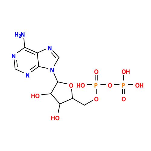 Mass: 427.201 Da / Num. of mol.: 5 / Source method: obtained synthetically / Formula: C10H15N5O10P2 / Comment: ADP, energy-carrying molecule*YM Mass: 427.201 Da / Num. of mol.: 5 / Source method: obtained synthetically / Formula: C10H15N5O10P2 / Comment: ADP, energy-carrying molecule*YM#4: Chemical | ChemComp-MG /   Mass: 24.305 Da / Num. of mol.: 5 / Source method: obtained synthetically / Formula: Mg Mass: 24.305 Da / Num. of mol.: 5 / Source method: obtained synthetically / Formula: MgHas ligand of interest | N | Has protein modification | Y | |
|---|
-Experimental details
-Experiment
| Experiment | Method: ELECTRON MICROSCOPY |
|---|---|
| EM experiment | Aggregation state: FILAMENT / 3D reconstruction method: single particle reconstruction |
- Sample preparation
| Component |
| ||||||||||||||||||||||||
|---|---|---|---|---|---|---|---|---|---|---|---|---|---|---|---|---|---|---|---|---|---|---|---|---|---|
| Molecular weight | Experimental value: NO | ||||||||||||||||||||||||
| Source (natural) |
| ||||||||||||||||||||||||
| Source (recombinant) | Organism: Escherichia coli (E. coli) | ||||||||||||||||||||||||
| Buffer solution | pH: 7.5 | ||||||||||||||||||||||||
| Specimen | Embedding applied: NO / Shadowing applied: NO / Staining applied: NO / Vitrification applied: YES | ||||||||||||||||||||||||
| Specimen support | Grid type: Quantifoil R1.2/1.3 | ||||||||||||||||||||||||
| Vitrification | Instrument: FEI VITROBOT MARK IV / Cryogen name: ETHANE / Humidity: 100 % / Chamber temperature: 277 K |
- Electron microscopy imaging
| Experimental equipment |  Model: Titan Krios / Image courtesy: FEI Company |
|---|---|
| Microscopy | Model: TFS KRIOS |
| Electron gun | Electron source:  FIELD EMISSION GUN / Accelerating voltage: 300 kV / Illumination mode: SPOT SCAN FIELD EMISSION GUN / Accelerating voltage: 300 kV / Illumination mode: SPOT SCAN |
| Electron lens | Mode: BRIGHT FIELD / Nominal magnification: 89000 X / Nominal defocus max: 2500 nm / Nominal defocus min: 500 nm |
| Specimen holder | Specimen holder model: FEI TITAN KRIOS AUTOGRID HOLDER |
| Image recording | Electron dose: 50 e/Å2 / Film or detector model: GATAN K3 (6k x 4k) |
- Processing
| EM software |
| ||||||||||||||||||||||||||||||||
|---|---|---|---|---|---|---|---|---|---|---|---|---|---|---|---|---|---|---|---|---|---|---|---|---|---|---|---|---|---|---|---|---|---|
| CTF correction | Type: PHASE FLIPPING AND AMPLITUDE CORRECTION | ||||||||||||||||||||||||||||||||
| 3D reconstruction | Resolution: 3.5 Å / Resolution method: FSC 0.143 CUT-OFF / Num. of particles: 29493 / Symmetry type: POINT | ||||||||||||||||||||||||||||||||
| Refinement | Highest resolution: 3.5 Å Stereochemistry target values: REAL-SPACE (WEIGHTED MAP SUM AT ATOM CENTERS) | ||||||||||||||||||||||||||||||||
| Refine LS restraints |
|
