 Movie
Movie Controller


+ Open data

- Basic information
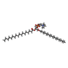
| Entry | Database: PDB / ID: 9pxf | |||||||||||||||||||||
|---|---|---|---|---|---|---|---|---|---|---|---|---|---|---|---|---|---|---|---|---|---|---|
| Title | Ammonia monooxygenase in native membranes from N. briensis | |||||||||||||||||||||
 Components Components | (Ammonia monooxygenase subunit ...) x 3 | |||||||||||||||||||||
 Keywords Keywords | OXIDOREDUCTASE / ammonia oxidation / copper enzyme / membrane protein / active site | |||||||||||||||||||||
| Function / homology |  Function and homology information Function and homology information | |||||||||||||||||||||
| Biological species |  Nitrosospira briensis (bacteria) Nitrosospira briensis (bacteria) | |||||||||||||||||||||
| Method | ELECTRON MICROSCOPY / single particle reconstruction / cryo EM / Resolution: 2.58 Å | |||||||||||||||||||||
 Authors Authors | Frank, F.J. / Rosenzweig, A.C. | |||||||||||||||||||||
| Funding support |  United States, 4items United States, 4items
| |||||||||||||||||||||
 Citation Citation | Journal: Chem Sci / Year: 2026 Title: Simultaneous occupancy of Cu and Cu in the ammonia monooxygenase active site. Authors: Frank J Tucci / Madeline B Ho / Aaron A B Turner / Lisa Y Stein / Brian M Hoffman / Amy C Rosenzweig /  Abstract: Ammonia monooxygenase (AMO), a copper-dependent membrane enzyme, catalyzes the first and rate-limiting step of nitrification: the oxidation of ammonia to hydroxylamine. Despite its central role in ...Ammonia monooxygenase (AMO), a copper-dependent membrane enzyme, catalyzes the first and rate-limiting step of nitrification: the oxidation of ammonia to hydroxylamine. Despite its central role in the global nitrogen cycle and its biotechnological relevance, structural characterization of AMO has lagged behind that of its homolog, particulate methane monooxygenase (pMMO), due to the slow growth rates of ammonia-oxidizing bacteria and the instability of AMO upon purification. Recent cryoEM studies of AMO and (Bath) pMMO in native membranes revealed new structural features, including two adjacent copper-binding sites in the transmembrane region, Cu and Cu, believed to constitute the active site. Although multiple structures were determined under various conditions, simultaneous occupancy of Cu and Cu was never observed, leaving their potential functional interplay unresolved. Here we report the 2.6 Å resolution cryoEM structure of AMO from C-128 in isolated native membranes. This structure reveals the first instance of simultaneous copper occupancy of the Cu and Cu sites, along with occupancy of the periplasmic Cu site. Electron paramagnetic resonance (EPR) spectroscopic data indicate that the Cu site is primarily occupied by Cu(ii), while Cu and Cu are primarily occupied by diamagnetic ions, presumably Cu(i). Notably, a lipid molecule is bound between the Cu and Cu sites, separating them by ∼8.0 Å. The results underscore the importance of studying these enzymes in their native environments across species to resolve conserved and divergent molecular features. | |||||||||||||||||||||
| History |
|
- Structure visualization
| Structure viewer | Molecule: MolmilJmol/JSmol |
|---|
- Downloads & links
-Download
| PDBx/mmCIF format | 9pxf.cif.gz | 570.2 KB | Display | PDBx/mmCIF format |
|---|---|---|---|---|
| PDB format | pdb9pxf.ent.gz | 464.2 KB | Display | PDB format |
| PDBx/mmJSON format | 9pxf.json.gz | Tree view | PDBx/mmJSON format | |
| Others |  Other downloads Other downloads |
-Validation report
| Arichive directory | https://data.pdbj.org/pub/pdb/validation_reports/px/9pxfftp://data.pdbj.org/pub/pdb/validation_reports/px/9pxf | HTTPS FTP |
|---|
-Related structure data
| Related structure data |  71966MC M: map data used to model this data C: citing same article ( |
|---|---|
| Similar structure data |
-Links
 PDBj
PDBj- Assembly
| Deposited unit | 
|
|---|---|
| 1 |
|
-Components
-Ammonia monooxygenase subunit ... , 3 types, 9 molecules GHIDEFABC
| #1: Protein | Mass: 29534.312 Da / Num. of mol.: 3 Source method: isolated from a genetically manipulated source Source: (gene. exp.) Nitrosospira briensis (bacteria) / Gene: SAMN05720354_1358 / Production host: Nitrosospira briensis (bacteria) / References: UniProt: A0A1G4ZCX6#2: Protein | Mass: 31288.760 Da / Num. of mol.: 3 Source method: isolated from a genetically manipulated source Source: (gene. exp.) Nitrosospira briensis (bacteria) / Gene: SAMN05216386_3025 / Production host: Nitrosospira briensis (bacteria) / References: UniProt: A0A1I5FI55#3: Protein | Mass: 42675.074 Da / Num. of mol.: 3 Source method: isolated from a genetically manipulated source Source: (gene. exp.) Nitrosospira briensis (bacteria) / Gene: SAMN05216386_3024 / Production host: Nitrosospira briensis (bacteria) / References: UniProt: A0A1I5FIF1 |
|---|
-Non-polymers , 3 types, 860 molecules 

| #4: Chemical | ChemComp-CU /  Mass: 63.546 Da / Num. of mol.: 9 / Source method: obtained synthetically / Formula: Cu / Feature type: SUBJECT OF INVESTIGATION Mass: 63.546 Da / Num. of mol.: 9 / Source method: obtained synthetically / Formula: Cu / Feature type: SUBJECT OF INVESTIGATION#5: Chemical |  Mass: 763.100 Da / Num. of mol.: 3 / Source method: obtained synthetically / Formula: C42H85NO8P / Comment: phospholipid*YM Mass: 763.100 Da / Num. of mol.: 3 / Source method: obtained synthetically / Formula: C42H85NO8P / Comment: phospholipid*YM#6: Water | ChemComp-HOH / | Mass: 18.015 Da / Num. of mol.: 848 / Source method: isolated from a natural source / Formula: H2O |
|---|
-Details
| Has ligand of interest | Y |
|---|---|
| Has protein modification | N |
-Experimental details
-Experiment
| Experiment | Method: ELECTRON MICROSCOPY |
|---|---|
| EM experiment | Aggregation state: 2D ARRAY / 3D reconstruction method: single particle reconstruction |
- Sample preparation
| Component | Name: Ammonia monooxygenase in native membranes from N. briensis Type: ORGANELLE OR CELLULAR COMPONENT / Entity ID: #1-#3 / Source: NATURAL |
|---|---|
| Source (natural) | Organism: Nitrosospira briensis (bacteria) |
| Buffer solution | pH: 7.2 |
| Specimen | Conc.: 3 mg/ml / Embedding applied: NO / Shadowing applied: NO / Staining applied: NO / Vitrification applied: YES |
| Vitrification | Cryogen name: ETHANE |
- Electron microscopy imaging
| Experimental equipment |  Model: Titan Krios / Image courtesy: FEI Company |
|---|---|
| Microscopy | Model: TFS KRIOS |
| Electron gun | Electron source:  FIELD EMISSION GUN / Accelerating voltage: 300 kV / Illumination mode: SPOT SCAN FIELD EMISSION GUN / Accelerating voltage: 300 kV / Illumination mode: SPOT SCAN |
| Electron lens | Mode: BRIGHT FIELD / Nominal defocus max: 2000 nm / Nominal defocus min: 600 nm |
| Image recording | Electron dose: 50 e/Å2 / Film or detector model: GATAN K3 (6k x 4k) |
- Processing
| EM software |
| ||||||||||||||||
|---|---|---|---|---|---|---|---|---|---|---|---|---|---|---|---|---|---|
| CTF correction | Type: PHASE FLIPPING AND AMPLITUDE CORRECTION | ||||||||||||||||
| 3D reconstruction | Resolution: 2.58 Å / Resolution method: FSC 0.143 CUT-OFF / Num. of particles: 212075 / Symmetry type: POINT |
