 Movie
Movie Controller

[English] 日本語
 Yorodumi
- PDB-9pwk: P21A Mutant of E. coli Dihydrofolate Reductase Complexed with Fol... -
+ Open data

- Basic information
| Entry | Database: PDB / ID: 9pwk | |||||||||
|---|---|---|---|---|---|---|---|---|---|---|
| Title | P21A Mutant of E. coli Dihydrofolate Reductase Complexed with Folate and Nicotinamide Adenine Dinucleotide Phosphate (oxidized form) | |||||||||
 Components Components | Dihydrofolate reductase | |||||||||
 Keywords Keywords | OXIDOREDUCTASE / P21A / DHFR | |||||||||
| Function / homology |  Function and homology information Function and homology informationmethotrexate binding / dihydrofolic acid binding / 10-formyltetrahydrofolate biosynthetic process / response to methotrexate / folic acid biosynthetic process / folic acid binding / NADP+ binding / dihydrofolate metabolic process / dihydrofolate reductase / dihydrofolate reductase activity ...methotrexate binding / dihydrofolic acid binding / 10-formyltetrahydrofolate biosynthetic process / response to methotrexate / folic acid biosynthetic process / folic acid binding / NADP+ binding / dihydrofolate metabolic process / dihydrofolate reductase / dihydrofolate reductase activity / folic acid metabolic process / tetrahydrofolate biosynthetic process / NADPH binding / one-carbon metabolic process / NADP binding / response to xenobiotic stimulus / response to antibiotic / cytosol Similarity search - Function | |||||||||
| Biological species |  | |||||||||
| Method |  X-RAY DIFFRACTION / SYNCHROTRON / MOLECULAR REPLACEMENT / Resolution: 1.51 Å X-RAY DIFFRACTION / SYNCHROTRON / MOLECULAR REPLACEMENT / Resolution: 1.51 Å | |||||||||
 Authors Authors | Fried, S.D.E. / Mathews, I.I. / Boxer, S.G. | |||||||||
| Funding support |  United States, 2items United States, 2items
| |||||||||
 Citation Citation | Journal: To Be Published Title: Role of Electrostatics in Hydride Transfer by Dihydrofolate Reductase Authors: Fried, S.D.E. / Mukherjee, S. / Boxer, S.G. | |||||||||
| History |
|
- Structure visualization
| Structure viewer | Molecule: MolmilJmol/JSmol |
|---|
- Downloads & links
-Download
| PDBx/mmCIF format | 9pwk.cif.gz | 54.1 KB | Display | PDBx/mmCIF format |
|---|---|---|---|---|
| PDB format | pdb9pwk.ent.gz | 35.6 KB | Display | PDB format |
| PDBx/mmJSON format | 9pwk.json.gz | Tree view | PDBx/mmJSON format | |
| Others |  Other downloads Other downloads |
-Validation report
| Arichive directory | https://data.pdbj.org/pub/pdb/validation_reports/pw/9pwkftp://data.pdbj.org/pub/pdb/validation_reports/pw/9pwk | HTTPS FTP |
|---|
-Related structure data
| Related structure data |  8ucxC  8vz4C  9ni0C  9njoC  9pwmC  9pxcC C: citing same article ( |
|---|---|
| Similar structure data |
-Links
 PDBj
PDBj
- Assembly
| Deposited unit | 
| ||||||||
|---|---|---|---|---|---|---|---|---|---|
| 1 |
| ||||||||
| Unit cell |
|
-Components
| #1: Protein | Mass: 17993.305 Da / Num. of mol.: 1 / Mutation: P21A Source method: isolated from a genetically manipulated source Source: (gene. exp.) |
|---|---|
| #2: Chemical | ChemComp-FOL / 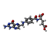  Mass: 441.397 Da / Num. of mol.: 1 / Source method: obtained synthetically / Formula: C19H19N7O6 / Feature type: SUBJECT OF INVESTIGATION Mass: 441.397 Da / Num. of mol.: 1 / Source method: obtained synthetically / Formula: C19H19N7O6 / Feature type: SUBJECT OF INVESTIGATION |
| #3: Chemical | ChemComp-NAP /   Mass: 743.405 Da / Num. of mol.: 1 / Source method: obtained synthetically / Formula: C21H28N7O17P3 / Feature type: SUBJECT OF INVESTIGATION Mass: 743.405 Da / Num. of mol.: 1 / Source method: obtained synthetically / Formula: C21H28N7O17P3 / Feature type: SUBJECT OF INVESTIGATION |
| #4: Chemical | ChemComp-CA /   Mass: 40.078 Da / Num. of mol.: 1 / Source method: obtained synthetically / Formula: Ca Mass: 40.078 Da / Num. of mol.: 1 / Source method: obtained synthetically / Formula: Ca |
| #5: Water | ChemComp-HOH /  Mass: 18.015 Da / Num. of mol.: 166 / Source method: isolated from a natural source / Formula: H2O Mass: 18.015 Da / Num. of mol.: 166 / Source method: isolated from a natural source / Formula: H2O |
| Has ligand of interest | Y |
| Has protein modification | N |
-Experimental details
-Experiment
| Experiment | Method: X-RAY DIFFRACTION / Number of used crystals: 1 |
|---|
- Sample preparation
| Crystal | Density Matthews: 2.12 Å3/Da / Density % sol: 42.09 % |
|---|---|
| Crystal grow | Temperature: 298 K / Method: vapor diffusion, hanging drop / pH: 8 / Details: PEG 6000, Calcium chloride, Tris |
-Data collection
| Diffraction | Mean temperature: 100 K / Serial crystal experiment: N |
|---|---|
| Diffraction source | Source: SYNCHROTRON / Site: SSRL / Beamline: BL12-1 / Wavelength: 0.9795 Å |
| Detector | Type: DECTRIS EIGER2 XE 16M / Detector: PIXEL / Date: Jul 9, 2025 |
| Radiation | Protocol: SINGLE WAVELENGTH / Monochromatic (M) / Laue (L): M / Scattering type: x-ray |
| Radiation wavelength | Wavelength: 0.9795 Å / Relative weight: 1 |
| Reflection | Resolution: 1.51→34.06 Å / Num. obs: 24773 / % possible obs: 99.6 % / Redundancy: 12.8 % / CC1/2: 0.969 / Rmerge(I) obs: 0.449 / Rpim(I) all: 0.131 / Rrim(I) all: 0.469 / Χ2: 0.87 / Net I/σ(I): 8.4 / Num. measured all: 316915 |
| Reflection shell | Resolution: 1.51→1.53 Å / % possible obs: 97.7 % / Redundancy: 11.9 % / Rmerge(I) obs: 7.405 / Num. measured all: 14301 / Num. unique obs: 1199 / CC1/2: 0.725 / Rpim(I) all: 2.149 / Rrim(I) all: 7.718 / Χ2: 1.19 / Net I/σ(I) obs: 1.9 |
- Processing
| Software |
| ||||||||||||||||||||||||||||||||||||||||||||||||||||||||||||||||||||||
|---|---|---|---|---|---|---|---|---|---|---|---|---|---|---|---|---|---|---|---|---|---|---|---|---|---|---|---|---|---|---|---|---|---|---|---|---|---|---|---|---|---|---|---|---|---|---|---|---|---|---|---|---|---|---|---|---|---|---|---|---|---|---|---|---|---|---|---|---|---|---|---|
| Refinement | Method to determine structure: MOLECULAR REPLACEMENT / Resolution: 1.51→32.75 Å / SU ML: 0.16 / Cross valid method: FREE R-VALUE / σ(F): 1.33 / Phase error: 23.66 / Stereochemistry target values: ML
| ||||||||||||||||||||||||||||||||||||||||||||||||||||||||||||||||||||||
| Solvent computation | Shrinkage radii: 0.9 Å / VDW probe radii: 1.1 Å / Solvent model: FLAT BULK SOLVENT MODEL | ||||||||||||||||||||||||||||||||||||||||||||||||||||||||||||||||||||||
| Refinement step | Cycle: LAST / Resolution: 1.51→32.75 Å
| ||||||||||||||||||||||||||||||||||||||||||||||||||||||||||||||||||||||
| Refine LS restraints |
| ||||||||||||||||||||||||||||||||||||||||||||||||||||||||||||||||||||||
| LS refinement shell |
|
