 Movie
Movie Controller


+ Open data

- Basic information
| Entry | Database: PDB / ID: 9p8u | ||||||||||||||||||||||||
|---|---|---|---|---|---|---|---|---|---|---|---|---|---|---|---|---|---|---|---|---|---|---|---|---|---|
| Title | Structure of CloA in complex with dGTP and p3diT | ||||||||||||||||||||||||
 Components Components |
| ||||||||||||||||||||||||
 Keywords Keywords | HYDROLASE / deoxynucleoside triphosphohydrolase | ||||||||||||||||||||||||
| Function / homology |  Function and homology information Function and homology information | ||||||||||||||||||||||||
| Biological species |  Salmonella enterica (bacteria) Salmonella enterica (bacteria)synthetic construct (others) | ||||||||||||||||||||||||
| Method | ELECTRON MICROSCOPY / single particle reconstruction / cryo EM / Resolution: 2.56 Å | ||||||||||||||||||||||||
 Authors Authors | Yamaguchi, S. / Fernandez, S.G. / Wassarman, D.R. / Luder, M. / Schwede, F. / Kranzusch, P.J. | ||||||||||||||||||||||||
| Funding support |  Japan, Japan,  France, France,  United States, 3items United States, 3items
| ||||||||||||||||||||||||
 Citation Citation | Journal: bioRxiv / Year: 2025 Title: Activating and inhibiting nucleotide signals coordinate bacterial anti-phage defense. Authors: Sonomi Yamaguchi / Samantha G Fernandez / Douglas R Wassarman / Marlen Lüders / Frank Schwede / Philip J Kranzusch /  Abstract: The cellular nucleotide pool is a major focal point of the host immune response to viral infection. Immune effector proteins that disrupt the nucleotide pool allow animal and bacterial cells to ...The cellular nucleotide pool is a major focal point of the host immune response to viral infection. Immune effector proteins that disrupt the nucleotide pool allow animal and bacterial cells to broadly restrict diverse viruses, but reduced nucleotide availability induces cellular toxicity and can limit host fitness(Ahmad et al., 1998; Goldstone et al., 2011; Hsueh et al., 2022; Itsko & Schaaper, 2014; Tal et al., 2022). Here we discover a bacterial anti-phage defense system named Clover that overcomes this tradeoff by encoding a deoxynucleoside triphosphohydrolase enzyme (CloA) that dynamically responds to both an activating phage cue and an inhibitory nucleotide immune signal produced by a partnering regulatory enzyme (CloB). Analysis of Clover phage restriction in cells and reconstitution of enzymatic function in vitro demonstrate that CloA is a dGTPase that responds to viral enzymes that increase cellular levels of dTTP. To restrain CloA activation in the absence of infection, we show that CloB synthesizes a dTTP-related inhibitory nucleotide signal p3diT (5'-triphosphothymidyl-3'5'-thymidine) that binds to CloA and suppresses activation. Cryo-EM structures of CloA in activated and suppressed states reveal how dTTP and p3diT control distinct allosteric sites and regulate effector function. Our results define how nucleotide signals coordinate both activation and inhibition of antiviral immunity and explain how cells balance defense and immune-mediated toxicity. | ||||||||||||||||||||||||
| History |
|
- Structure visualization
| Structure viewer | Molecule: MolmilJmol/JSmol |
|---|
- Downloads & links
-Download
| PDBx/mmCIF format | 9p8u.cif.gz | 918.5 KB | Display | PDBx/mmCIF format |
|---|---|---|---|---|
| PDB format | pdb9p8u.ent.gz | 613.2 KB | Display | PDB format |
| PDBx/mmJSON format | 9p8u.json.gz | Tree view | PDBx/mmJSON format | |
| Others |  Other downloads Other downloads |
-Validation report
| Arichive directory | https://data.pdbj.org/pub/pdb/validation_reports/p8/9p8uftp://data.pdbj.org/pub/pdb/validation_reports/p8/9p8u | HTTPS FTP |
|---|
-Related structure data
| Related structure data |  71389MC  9p8sC  9p8tC  9p8vC  9p8wC M: map data used to model this data C: citing same article ( |
|---|---|
| Similar structure data |


-Links
 PDBj
PDBj
- Assembly
| Deposited unit | 
|
|---|---|
| 1 |
|
-Components
| #1: Protein | Mass: 54257.172 Da / Num. of mol.: 8 Source method: isolated from a genetically manipulated source Source: (gene. exp.) Salmonella enterica (bacteria) / Gene: dgt, ECD07_17535, EIW74_15545, GB147_17355 / Production host: #2: DNA chain | Mass: 723.388 Da / Num. of mol.: 8 / Source method: obtained synthetically / Source: (synth.) synthetic construct (others) #3: Chemical | ChemComp-DGT / 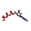  Mass: 507.181 Da / Num. of mol.: 8 / Source method: obtained synthetically / Formula: C10H16N5O13P3 / Feature type: SUBJECT OF INVESTIGATION Mass: 507.181 Da / Num. of mol.: 8 / Source method: obtained synthetically / Formula: C10H16N5O13P3 / Feature type: SUBJECT OF INVESTIGATION#4: Chemical | ChemComp-MG /   Mass: 24.305 Da / Num. of mol.: 8 / Source method: obtained synthetically / Formula: Mg Mass: 24.305 Da / Num. of mol.: 8 / Source method: obtained synthetically / Formula: MgHas ligand of interest | Y | Has protein modification | N | |
|---|
-Experimental details
-Experiment
| Experiment | Method: ELECTRON MICROSCOPY |
|---|---|
| EM experiment | Aggregation state: PARTICLE / 3D reconstruction method: single particle reconstruction |
- Sample preparation
| Component | Name: Octameric complex of CloA in complex with dGTP and p3diT Type: COMPLEX / Entity ID: #1-#2 / Source: MULTIPLE SOURCES |
|---|---|
| Source (natural) | Organism: Salmonella enterica (bacteria) |
| Source (recombinant) | Organism: |
| Buffer solution | pH: 7.4 |
| Specimen | Embedding applied: NO / Shadowing applied: NO / Staining applied: NO / Vitrification applied: YES |
| Vitrification | Cryogen name: ETHANE |
- Electron microscopy imaging
| Experimental equipment |  Model: Talos Arctica / Image courtesy: FEI Company |
|---|---|
| Microscopy | Model: FEI TALOS ARCTICA |
| Electron gun | Electron source:  FIELD EMISSION GUN / Accelerating voltage: 200 kV / Illumination mode: FLOOD BEAM FIELD EMISSION GUN / Accelerating voltage: 200 kV / Illumination mode: FLOOD BEAM |
| Electron lens | Mode: BRIGHT FIELD / Nominal defocus max: 2200 nm / Nominal defocus min: 900 nm |
| Image recording | Electron dose: 50.24 e/Å2 / Film or detector model: GATAN K3 (6k x 4k) |
- Processing
| EM software |
| ||||||||||||||||||||||||
|---|---|---|---|---|---|---|---|---|---|---|---|---|---|---|---|---|---|---|---|---|---|---|---|---|---|
| CTF correction | Type: PHASE FLIPPING AND AMPLITUDE CORRECTION | ||||||||||||||||||||||||
| 3D reconstruction | Resolution: 2.56 Å / Resolution method: FSC 0.143 CUT-OFF / Num. of particles: 1498407 / Symmetry type: POINT | ||||||||||||||||||||||||
| Refinement | Cross valid method: NONE Stereochemistry target values: GeoStd + Monomer Library + CDL v1.2 | ||||||||||||||||||||||||
| Displacement parameters | Biso mean: 127.9 Å2 | ||||||||||||||||||||||||
| Refine LS restraints |
|
