 Movie
Movie Controller

[English] 日本語
 Yorodumi
- PDB-9p8a: NTSR1-G11-NTS(8-13) GTP-Bound Complex in the Canonical, AHD Close... -
+ Open data

- Basic information
| Entry | Database: PDB / ID: 9p8a | ||||||||||||||||||||||||
|---|---|---|---|---|---|---|---|---|---|---|---|---|---|---|---|---|---|---|---|---|---|---|---|---|---|
| Title | NTSR1-G11-NTS(8-13) GTP-Bound Complex in the Canonical, AHD Closed State 3DVA Separated 2 (C-Closed*-GTP) | ||||||||||||||||||||||||
 Components Components |
| ||||||||||||||||||||||||
 Keywords Keywords | SIGNALING PROTEIN / Complex / Agonist | ||||||||||||||||||||||||
| Function / homology |  Function and homology information Function and homology informationregulation of melanocyte differentiation / phospholipase C-activating G protein-coupled acetylcholine receptor signaling pathway / endothelin receptor signaling pathway / Fatty Acids bound to GPR40 (FFAR1) regulate insulin secretion / Acetylcholine regulates insulin secretion / developmental pigmentation / phospholipase C-activating dopamine receptor signaling pathway / cellular response to pH / PLC beta mediated events / entrainment of circadian clock ...regulation of melanocyte differentiation / phospholipase C-activating G protein-coupled acetylcholine receptor signaling pathway / endothelin receptor signaling pathway / Fatty Acids bound to GPR40 (FFAR1) regulate insulin secretion / Acetylcholine regulates insulin secretion / developmental pigmentation / phospholipase C-activating dopamine receptor signaling pathway / cellular response to pH / PLC beta mediated events / entrainment of circadian clock / cranial skeletal system development / ligand-gated ion channel signaling pathway / phototransduction, visible light / action potential / photoreceptor outer segment / enzyme regulator activity / Turbulent (oscillatory, disturbed) flow shear stress activates signaling by PIEZO1 and integrins in endothelial cells / skeletal system development / G protein-coupled receptor binding / regulation of blood pressure / G-protein beta/gamma-subunit complex binding / adenylate cyclase-modulating G protein-coupled receptor signaling pathway / positive regulation of insulin secretion / Olfactory Signaling Pathway / Activation of the phototransduction cascade / G protein-coupled acetylcholine receptor signaling pathway / G beta:gamma signalling through PLC beta / Presynaptic function of Kainate receptors / Thromboxane signalling through TP receptor / Activation of G protein gated Potassium channels / Inhibition of voltage gated Ca2+ channels via Gbeta/gamma subunits / G-protein activation / Glucagon signaling in metabolic regulation / G beta:gamma signalling through CDC42 / Prostacyclin signalling through prostacyclin receptor / Synthesis, secretion, and inactivation of Glucagon-like Peptide-1 (GLP-1) / G beta:gamma signalling through BTK / photoreceptor disc membrane / ADP signalling through P2Y purinoceptor 12 / Glucagon-type ligand receptors / Sensory perception of sweet, bitter, and umami (glutamate) taste / Adrenaline,noradrenaline inhibits insulin secretion / Vasopressin regulates renal water homeostasis via Aquaporins / Glucagon-like Peptide-1 (GLP1) regulates insulin secretion / G alpha (z) signalling events / cellular response to catecholamine stimulus / ADP signalling through P2Y purinoceptor 1 / G beta:gamma signalling through PI3Kgamma / ADORA2B mediated anti-inflammatory cytokines production / adenylate cyclase-activating dopamine receptor signaling pathway / Cooperation of PDCL (PhLP1) and TRiC/CCT in G-protein beta folding / GPER1 signaling / cellular response to prostaglandin E stimulus / heterotrimeric G-protein complex / G alpha (12/13) signalling events / Inactivation, recovery and regulation of the phototransduction cascade / G-protein beta-subunit binding / extracellular vesicle / heart development / sensory perception of taste / Thrombin signalling through proteinase activated receptors (PARs) / signaling receptor complex adaptor activity / retina development in camera-type eye / fibroblast proliferation / GTPase binding / G protein activity / Ca2+ pathway / High laminar flow shear stress activates signaling by PIEZO1 and PECAM1:CDH5:KDR in endothelial cells / G alpha (i) signalling events / G alpha (s) signalling events / G alpha (q) signalling events / phospholipase C-activating G protein-coupled receptor signaling pathway / Hydrolases; Acting on acid anhydrides; Acting on GTP to facilitate cellular and subcellular movement / Ras protein signal transduction / Extra-nuclear estrogen signaling / cell population proliferation / G protein-coupled receptor signaling pathway / lysosomal membrane / GTPase activity / synapse / GTP binding / protein-containing complex binding / signal transduction / extracellular exosome / membrane / metal ion binding / plasma membrane / cytoplasm / cytosol Similarity search - Function | ||||||||||||||||||||||||
| Biological species |  Homo sapiens (human) Homo sapiens (human) | ||||||||||||||||||||||||
| Method | ELECTRON MICROSCOPY / single particle reconstruction / cryo EM / Resolution: 3.7 Å | ||||||||||||||||||||||||
 Authors Authors | Robertson, M.J. | ||||||||||||||||||||||||
| Funding support |  United States, 2items United States, 2items
| ||||||||||||||||||||||||
 Citation Citation | Journal: Nature / Year: 2026 Title: Snapshots of the dynamic basis of NTSR1 G protein subtype promiscuity. Authors: Alina A Vo / Arnab Modak / Sumin Lu / Scott C Blanchard / Nevin A Lambert / Michael J Robertson / Abstract: G-protein-coupled receptors (GPCRs) are capable of signalling through four families of G protein α subunits. Although hundreds of nucleotide-free GPCR-G protein complex structures have been solved, ...G-protein-coupled receptors (GPCRs) are capable of signalling through four families of G protein α subunits. Although hundreds of nucleotide-free GPCR-G protein complex structures have been solved, the mechanism of G protein subtype selectivity remains poorly understood, with recent studies suggesting a role for dynamic nucleotide-bound intermediate states. Here we use time-resolved cryo-electron microscopy to visualize the GTP-induced activation of Gαβγ and Gαβγ heterotrimers bound to the neurotensin receptor 1 (NTSR1), which has been demonstrated to be highly promiscuous in G protein coupling and to possess unusual conformations in the nucleotide-free complex. We resolve ensembles of states along the G protein activation pathway, with differences in the structures and their relative populations between Gα and Gα. Structural analysis reveals a key role for several motifs, including intracellular loop 2 (ICL2) and ICL3, in stabilizing the observed intermediate states. Our results are supported by molecular dynamics simulations and kinetic bioluminescence resonance energy transfer experiments, which reveal that the stability of these intermediate states and the signalling of various G proteins are correlated with ICL2 and ICL3 sequences. Single-molecule fluorescence assays of GTP-induced NTSR1-G protein complex dissociation reveal that NTSR1 is liberated significantly faster from Gα, consistent with the relative lack of stable Gα-GTP intermediate states compared with Gα. These findings highlight that transient intermediate-state complexes along the G protein activation pathway have an important role in G protein selection that cannot be explained by nucleotide-free states alone. | ||||||||||||||||||||||||
| History |
|
- Structure visualization
| Structure viewer | Molecule: MolmilJmol/JSmol |
|---|
- Downloads & links
-Download
| PDBx/mmCIF format | 9p8a.cif.gz | 146.1 KB | Display | PDBx/mmCIF format |
|---|---|---|---|---|
| PDB format | pdb9p8a.ent.gz | 103 KB | Display | PDB format |
| PDBx/mmJSON format | 9p8a.json.gz | Tree view | PDBx/mmJSON format | |
| Others |  Other downloads Other downloads |
-Validation report
| Arichive directory | https://data.pdbj.org/pub/pdb/validation_reports/p8/9p8aftp://data.pdbj.org/pub/pdb/validation_reports/p8/9p8a | HTTPS FTP |
|---|
-Related structure data
| Related structure data |  71370MC  9p7zC  9p80C  9p81C  9p82C  9p83C  9p84C  9p85C  9p86C 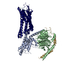 9p87C  9p88C  9p89C M: map data used to model this data C: citing same article ( |
|---|---|
| Similar structure data |


-Links
 PDBj
PDBj


- Assembly
| Deposited unit | 
|
|---|---|
| 1 |
|
-Components
| #1: Protein | Mass: 42177.254 Da / Num. of mol.: 1 Source method: isolated from a genetically manipulated source Source: (gene. exp.) Homo sapiens (human) / Gene: GNA11, GA11 / Production host:  Trichoplusia ni (cabbage looper) Trichoplusia ni (cabbage looper)References: UniProt: P29992, Hydrolases; Acting on acid anhydrides; Acting on GTP to facilitate cellular and subcellular movement |
|---|---|
| #2: Protein | Mass: 37671.102 Da / Num. of mol.: 1 Source method: isolated from a genetically manipulated source Source: (gene. exp.) Homo sapiens (human) / Gene: GNB1 / Production host: Trichoplusia ni (cabbage looper) / References: UniProt: P62873 |
| #3: Protein | Mass: 7861.143 Da / Num. of mol.: 1 Source method: isolated from a genetically manipulated source Source: (gene. exp.) Homo sapiens (human) / Gene: GNG2 / Production host: Trichoplusia ni (cabbage looper) / References: UniProt: P59768 |
| #4: Chemical | ChemComp-GTP /   Mass: 523.180 Da / Num. of mol.: 1 / Source method: isolated from a natural source / Formula: C10H16N5O14P3 / Feature type: SUBJECT OF INVESTIGATION / Comment: GTP, energy-carrying molecule*YM Mass: 523.180 Da / Num. of mol.: 1 / Source method: isolated from a natural source / Formula: C10H16N5O14P3 / Feature type: SUBJECT OF INVESTIGATION / Comment: GTP, energy-carrying molecule*YM |
| #5: Water | ChemComp-HOH /  Mass: 18.015 Da / Num. of mol.: 4 / Source method: isolated from a natural source / Formula: H2O Mass: 18.015 Da / Num. of mol.: 4 / Source method: isolated from a natural source / Formula: H2O |
| Has ligand of interest | Y |
| Has protein modification | N |
-Experimental details
-Experiment
| Experiment | Method: ELECTRON MICROSCOPY |
|---|---|
| EM experiment | Aggregation state: PARTICLE / 3D reconstruction method: single particle reconstruction |
- Sample preparation
| Component | Name: NTSR1-Gi-NTS(8-13) Complex in the Canonical, AHD Open State Type: COMPLEX / Entity ID: #1-#3 / Source: RECOMBINANT |
|---|---|
| Source (natural) | Organism: Homo sapiens (human) |
| Source (recombinant) | Organism:  Spodoptera frugiperda (fall armyworm) Spodoptera frugiperda (fall armyworm) |
| Buffer solution | pH: 7.5 |
| Specimen | Embedding applied: NO / Shadowing applied: NO / Staining applied: NO / Vitrification applied: YES |
| Vitrification | Cryogen name: ETHANE |
- Electron microscopy imaging
| Experimental equipment |  Model: Titan Krios / Image courtesy: FEI Company |
|---|---|
| Microscopy | Model: TFS KRIOS |
| Electron gun | Electron source:  FIELD EMISSION GUN / Accelerating voltage: 300 kV / Illumination mode: FLOOD BEAM FIELD EMISSION GUN / Accelerating voltage: 300 kV / Illumination mode: FLOOD BEAM |
| Electron lens | Mode: BRIGHT FIELD / Nominal defocus max: 1400 nm / Nominal defocus min: 600 nm |
| Image recording | Electron dose: 55 e/Å2 / Film or detector model: GATAN K3 BIOQUANTUM (6k x 4k) |
- Processing
| EM software |
| ||||||||||||||||||||||||
|---|---|---|---|---|---|---|---|---|---|---|---|---|---|---|---|---|---|---|---|---|---|---|---|---|---|
| CTF correction | Type: PHASE FLIPPING AND AMPLITUDE CORRECTION | ||||||||||||||||||||||||
| 3D reconstruction | Resolution: 3.7 Å / Resolution method: FSC 0.143 CUT-OFF / Num. of particles: 73764 / Symmetry type: POINT | ||||||||||||||||||||||||
| Refine LS restraints |
|
