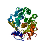
Movie
Movie Controller

[English] 日本語
 Yorodumi
- PDB-9oyy: MicroED structure of proteinase K from microcrystals frozen by tr... -
+ Open data

- Basic information
| Entry | Database: PDB / ID: 9oyy | ||||||
|---|---|---|---|---|---|---|---|
| Title | MicroED structure of proteinase K from microcrystals frozen by traditional cryoEM methods | ||||||
 Components Components | Proteinase K | ||||||
 Keywords Keywords | HYDROLASE / enzyme / MicroED | ||||||
| Function / homology |  Function and homology information Function and homology informationpeptidase K / serine-type endopeptidase activity / proteolysis / extracellular region / metal ion binding Similarity search - Function | ||||||
| Biological species |  Parengyodontium album (fungus) Parengyodontium album (fungus) | ||||||
| Method | ELECTRON CRYSTALLOGRAPHY / electron crystallography /  MOLECULAR REPLACEMENT / cryo EM / Resolution: 2.2 Å MOLECULAR REPLACEMENT / cryo EM / Resolution: 2.2 Å | ||||||
 Authors Authors | Vlahakis, N. / Summers, J.A. / Rodriguez, J.A. / Dahlberg, P. / Wakatsuki, S. | ||||||
| Funding support |  United States, 1items United States, 1items
| ||||||
 Citation Citation | Journal: To Be Published Title: Mix-and-spray grid preparation for time-resolved MicroED Authors: Summers, J.A. / Vlahakis, N.W. / Zielinski, K.A. / Uttormark, S. / Dolamore, C. / Antolini, C. / Wilson, M.A. / Pollack, L. / Rodriguez, J.A. / Dahlberg, P.D. / Wakatsuki, S. | ||||||
| History |
|
- Structure visualization
| Structure viewer | Molecule: MolmilJmol/JSmol |
|---|
- Downloads & links
-Download
| PDBx/mmCIF format | 9oyy.cif.gz | 113.3 KB | Display | PDBx/mmCIF format |
|---|---|---|---|---|
| PDB format | pdb9oyy.ent.gz | 85.2 KB | Display | PDB format |
| PDBx/mmJSON format | 9oyy.json.gz | Tree view | PDBx/mmJSON format | |
| Others |  Other downloads Other downloads |
-Validation report
| Arichive directory | https://data.pdbj.org/pub/pdb/validation_reports/oy/9oyyftp://data.pdbj.org/pub/pdb/validation_reports/oy/9oyy | HTTPS FTP |
|---|
-Related structure data

-Links
 PDBj
PDBj

- Assembly
| Deposited unit | 
| ||||||||
|---|---|---|---|---|---|---|---|---|---|
| 1 | 
| ||||||||
| 2 | 
| ||||||||
| Unit cell |
|
-Components
| #1: Protein | Mass: 28958.791 Da / Num. of mol.: 2 / Source method: isolated from a natural source / Source: (natural) Parengyodontium album (fungus) / References: UniProt: P06873, peptidase K#2: Chemical | ChemComp-CA /   Mass: 40.078 Da / Num. of mol.: 4 / Source method: obtained synthetically / Formula: Ca Mass: 40.078 Da / Num. of mol.: 4 / Source method: obtained synthetically / Formula: Ca#3: Water | ChemComp-HOH / |  Mass: 18.015 Da / Num. of mol.: 18 / Source method: isolated from a natural source / Formula: H2O Mass: 18.015 Da / Num. of mol.: 18 / Source method: isolated from a natural source / Formula: H2OHas ligand of interest | N | Has protein modification | Y | |
|---|
-Experimental details
-Experiment
| Experiment | Method: ELECTRON CRYSTALLOGRAPHY |
|---|---|
| EM experiment | Aggregation state: 3D ARRAY / 3D reconstruction method: electron crystallography |
- Sample preparation
| Component | Name: Proteinase K / Type: COMPLEX Details: Crystals formed from 50 mg/mL proteinase K mixed 1:1 with reservoir solution (0.1 M Tris HCl pH 8.0, 1.2 M ammonium sulfate) Entity ID: #1 / Source: NATURAL |
|---|---|
| Source (natural) | Organism: Parengyodontium album (fungus) |
| EM crystal formation | Details: Crystals formed from 50 mg/mL proteinase K mixed 1:1 with reservoir solution (0.1 M Tris HCl pH 8.0, 1.2 M ammonium sulfate) Temperature: 293 K |
| Buffer solution | pH: 8 |
| Specimen | Embedding applied: NO / Shadowing applied: NO / Staining applied: NO / Vitrification applied: YES |
| Vitrification | Cryogen name: ETHANE |
-Data collection
| Experimental equipment |  Model: Titan Krios / Image courtesy: FEI Company |
|---|---|
| Microscopy | Model: TFS KRIOS |
| Electron gun | Electron source: FIELD EMISSION GUN / Accelerating voltage: 300 kV / Illumination mode: FLOOD BEAM |
| Electron lens | Mode: DIFFRACTION / Nominal defocus max: 0 nm / Nominal defocus min: 0 nm |
| Specimen holder | Temperature (max): 100 K / Temperature (min): 100 K |
| Image recording | Electron dose: 0.15 e/Å2 / Film or detector model: FEI CETA (4k x 4k) |
| EM diffraction shell | Resolution: 2.2→2.3 Å / Fourier space coverage: 59.55 % / Multiplicity: 3.29 / Num. of structure factors: 1409 / Phase residual: 33.9 ° |
| EM diffraction stats | Fourier space coverage: 79.86 % / High resolution: 2.2 Å / Num. of intensities measured: 120010 / Num. of structure factors: 36443 / Phase error rejection criteria: not provided / Rmerge: 0.266 |
- Processing
| EM 3D crystal entity | ∠α: 90 ° / ∠β: 90 ° / ∠γ: 90 ° / A: 38.3 Å / B: 129.1 Å / C: 47.7 Å / Space group name: P21 / Space group num: 4 | ||||||||||||||||||||||||||||||||||||||||||||||||||||||||||||||||||||||||||||||||||||||||||||||||||||||||||
|---|---|---|---|---|---|---|---|---|---|---|---|---|---|---|---|---|---|---|---|---|---|---|---|---|---|---|---|---|---|---|---|---|---|---|---|---|---|---|---|---|---|---|---|---|---|---|---|---|---|---|---|---|---|---|---|---|---|---|---|---|---|---|---|---|---|---|---|---|---|---|---|---|---|---|---|---|---|---|---|---|---|---|---|---|---|---|---|---|---|---|---|---|---|---|---|---|---|---|---|---|---|---|---|---|---|---|---|
| CTF correction | Type: NONE | ||||||||||||||||||||||||||||||||||||||||||||||||||||||||||||||||||||||||||||||||||||||||||||||||||||||||||
| 3D reconstruction | Resolution: 2.2 Å / Resolution method: DIFFRACTION PATTERN/LAYERLINES / Symmetry type: 3D CRYSTAL | ||||||||||||||||||||||||||||||||||||||||||||||||||||||||||||||||||||||||||||||||||||||||||||||||||||||||||
| Atomic model building | PDB-ID: 2ID8 Accession code: 2ID8 / Source name: PDB / Type: experimental model | ||||||||||||||||||||||||||||||||||||||||||||||||||||||||||||||||||||||||||||||||||||||||||||||||||||||||||
| Refinement | Method to determine structure: MOLECULAR REPLACEMENT / Resolution: 2.2→32.94 Å / Cor.coef. Fo:Fc: 0.911 / Cor.coef. Fo:Fc free: 0.896 / SU B: 6.33 / SU ML: 0.162 / Cross valid method: THROUGHOUT / ESU R: 0.281 / ESU R Free: 0.06 / Stereochemistry target values: MAXIMUM LIKELIHOOD / Details: HYDROGENS HAVE BEEN ADDED IN THE RIDING POSITIONS
| ||||||||||||||||||||||||||||||||||||||||||||||||||||||||||||||||||||||||||||||||||||||||||||||||||||||||||
| Solvent computation | Ion probe radii: 0.8 Å / Shrinkage radii: 0.8 Å / VDW probe radii: 1.2 Å / Solvent model: MASK | ||||||||||||||||||||||||||||||||||||||||||||||||||||||||||||||||||||||||||||||||||||||||||||||||||||||||||
| Displacement parameters | Biso mean: 25.078 Å2
| ||||||||||||||||||||||||||||||||||||||||||||||||||||||||||||||||||||||||||||||||||||||||||||||||||||||||||
| Refinement step | Cycle: 1 / Total: 4092 | ||||||||||||||||||||||||||||||||||||||||||||||||||||||||||||||||||||||||||||||||||||||||||||||||||||||||||
| Refine LS restraints |
|