[English] 日本語
Yorodumi
- PDB-9ovx: CRYSTAL STRUCTURE OF UBIQUITIN K27M MUTANT -

+
Open data


ID or keywords:

Loading...

-
Basic information

Entry
Database: PDB / ID: 9ovx
TitleCRYSTAL STRUCTURE OF UBIQUITIN K27M MUTANT
ComponentsUbiquitin
KeywordsSIGNALING PROTEIN / protein degradation / protein modification / proteasome-targeting / proteolytic pathway / PROTEIN TRANSPORT
Function / homology
Function and homology information


Maturation of protein E / Maturation of protein E / ER Quality Control Compartment (ERQC) / Myoclonic epilepsy of Lafora / FLT3 signaling by CBL mutants / IRAK2 mediated activation of TAK1 complex / Alpha-protein kinase 1 signaling pathway / Glycogen synthesis / IRAK1 recruits IKK complex / IRAK1 recruits IKK complex upon TLR7/8 or 9 stimulation ...Maturation of protein E / Maturation of protein E / ER Quality Control Compartment (ERQC) / Myoclonic epilepsy of Lafora / FLT3 signaling by CBL mutants / IRAK2 mediated activation of TAK1 complex / Alpha-protein kinase 1 signaling pathway / Glycogen synthesis / IRAK1 recruits IKK complex / IRAK1 recruits IKK complex upon TLR7/8 or 9 stimulation / Prevention of phagosomal-lysosomal fusion / Endosomal Sorting Complex Required For Transport (ESCRT) / Membrane binding and targetting of GAG proteins / Regulation of TBK1, IKKε (IKBKE)-mediated activation of IRF3, IRF7 / Negative regulation of FLT3 / PTK6 Regulates RTKs and Their Effectors AKT1 and DOK1 / Regulation of TBK1, IKKε-mediated activation of IRF3, IRF7 upon TLR3 ligation / IRAK2 mediated activation of TAK1 complex upon TLR7/8 or 9 stimulation / Constitutive Signaling by NOTCH1 HD Domain Mutants / NOTCH2 Activation and Transmission of Signal to the Nucleus / TICAM1,TRAF6-dependent induction of TAK1 complex / TICAM1-dependent activation of IRF3/IRF7 / APC/C:Cdc20 mediated degradation of Cyclin B / Downregulation of ERBB4 signaling / APC-Cdc20 mediated degradation of Nek2A / Regulation of FZD by ubiquitination / p75NTR recruits signalling complexes / InlA-mediated entry of Listeria monocytogenes into host cells / TRAF6 mediated IRF7 activation in TLR7/8 or 9 signaling / NF-kB is activated and signals survival / TRAF6-mediated induction of TAK1 complex within TLR4 complex / Regulation of pyruvate metabolism / Pexophagy / Downregulation of ERBB2:ERBB3 signaling / Regulation of innate immune responses to cytosolic DNA / NRIF signals cell death from the nucleus / Regulation of PTEN localization / VLDLR internalisation and degradation / Activated NOTCH1 Transmits Signal to the Nucleus / Synthesis of active ubiquitin: roles of E1 and E2 enzymes / Translesion synthesis by REV1 / TICAM1, RIP1-mediated IKK complex recruitment / Regulation of BACH1 activity / Translesion synthesis by POLK / JNK (c-Jun kinases) phosphorylation and activation mediated by activated human TAK1 / InlB-mediated entry of Listeria monocytogenes into host cell / MAP3K8 (TPL2)-dependent MAPK1/3 activation / Activation of IRF3, IRF7 mediated by TBK1, IKKε (IKBKE) / Downregulation of TGF-beta receptor signaling / Translesion synthesis by POLI / Josephin domain DUBs / Gap-filling DNA repair synthesis and ligation in GG-NER / IKK complex recruitment mediated by RIP1 / PINK1-PRKN Mediated Mitophagy / TGF-beta receptor signaling in EMT (epithelial to mesenchymal transition) / TNFR1-induced NF-kappa-B signaling pathway / Regulation of activated PAK-2p34 by proteasome mediated degradation / TCF dependent signaling in response to WNT / Regulation of NF-kappa B signaling / activated TAK1 mediates p38 MAPK activation / Autodegradation of Cdh1 by Cdh1:APC/C / APC/C:Cdc20 mediated degradation of Securin / NOTCH3 Activation and Transmission of Signal to the Nucleus / Regulation of signaling by CBL / Negative regulators of DDX58/IFIH1 signaling / N-glycan trimming in the ER and Calnexin/Calreticulin cycle / Asymmetric localization of PCP proteins / Fanconi Anemia Pathway / Negative regulation of FGFR3 signaling / Ubiquitin-dependent degradation of Cyclin D / Peroxisomal protein import / Deactivation of the beta-catenin transactivating complex / SCF-beta-TrCP mediated degradation of Emi1 / NIK-->noncanonical NF-kB signaling / Stabilization of p53 / AUF1 (hnRNP D0) binds and destabilizes mRNA / TNFR2 non-canonical NF-kB pathway / Negative regulation of FGFR2 signaling / Negative regulation of FGFR4 signaling / Downregulation of SMAD2/3:SMAD4 transcriptional activity / Negative regulation of FGFR1 signaling / Termination of translesion DNA synthesis / Assembly of the pre-replicative complex / EGFR downregulation / Vpu mediated degradation of CD4 / Regulation of TNFR1 signaling / Cdc20:Phospho-APC/C mediated degradation of Cyclin A / Assembly Of The HIV Virion / SMAD2/SMAD3:SMAD4 heterotrimer regulates transcription / Dectin-1 mediated noncanonical NF-kB signaling / Degradation of DVL / Degradation of AXIN / Degradation of CRY and PER proteins / Spry regulation of FGF signaling / Late endosomal microautophagy / Hh mutants are degraded by ERAD / Iron uptake and transport / Activation of NF-kappaB in B cells / NOD1/2 Signaling Pathway / G2/M Checkpoints
Similarity search - Function
: / Ubiquitin domain signature. / Ubiquitin conserved site / Ubiquitin domain / Ubiquitin family / Ubiquitin homologues / Ubiquitin domain profile. / Ubiquitin-like domain / Ubiquitin-like domain superfamily
Similarity search - Domain/homology
Biological speciesHomo sapiens (human)
MethodX-RAY DIFFRACTION / SYNCHROTRON / SAD / Resolution: 1.5 Å
AuthorsLubkowski, J. / Wentz, B.G. / Fushman, D. / Gardiner, C.
Funding support United States, 1items
OrganizationGrant numberCountry
National Institutes of Health/National Institute of General Medical Sciences (NIH/NIGMS)GM065334 United States
CitationJournal: To Be Published
Title: The impact of Lysine-27 mutation on ubiquitin structure, dynamics, and receptor recognition
Authors: Gardiner, C. / Wentz, B.G. / Lubkowski, J. / Fushman, D.
History
DepositionJun 1, 2025Deposition site: RCSB / Processing site: RCSB
Revision 1.0Apr 15, 2026Provider: repository / Type: Initial release

-
Structure visualization

Structure viewerMolecule:
MolmilJmol/JSmol

Downloads & links

-
Assembly

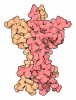
Deposited unit
A: Ubiquitin
hetero molecules


Theoretical massNumber of molelcules
Total (without water)9,46613
Polymers8,5791
Non-polymers88712
Water1,35175
1


  • Idetical with deposited unit
  • defined by author
  • Evidence: homology
TypeNameSymmetry operationNumber
identity operation1_555x,y,z1
Unit cell
Length a, b, c (Å)144.574, 144.574, 144.574
Angle α, β, γ (deg.)90.00, 90.00, 90.00
Int Tables number209
Space group name H-MF432
Components on special symmetry positions
IDModelComponents
11A-101-

CD

21A-105-

CD

31A-245-

HOH

41A-258-

HOH

51A-269-

HOH

-
Components

#1: Protein Ubiquitin


Mass: 8578.847 Da / Num. of mol.: 1 / Mutation: K27M
Source method: isolated from a genetically manipulated source
Details: Differs from wild type ubiquitin by the mutation Lys -> Met in position 27. Last four residues were disordered in the crystal structure.
Source: (gene. exp.) Homo sapiens (human) / Gene: UBC / Production host: Escherichia coli BL21(DE3) (bacteria) / Strain (production host): BL21(DE3) / References: UniProt: P0CG48
#2: Chemical
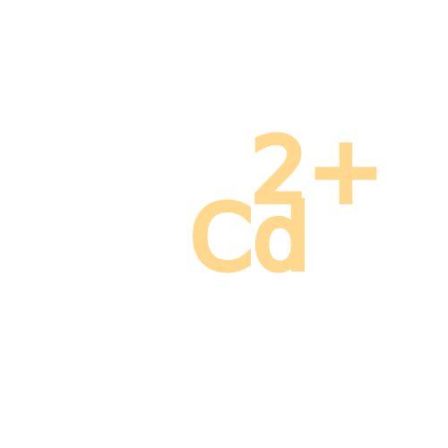
ChemComp-CD / CADMIUM ION


Mass: 112.411 Da / Num. of mol.: 6 / Source method: obtained synthetically / Formula: Cd
#3: Chemical
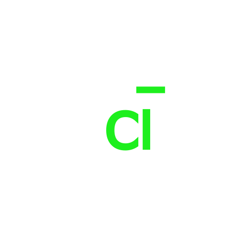
ChemComp-CL / CHLORIDE ION


Mass: 35.453 Da / Num. of mol.: 6 / Source method: obtained synthetically / Formula: Cl
#4: Water ChemComp-HOH / water


Mass: 18.015 Da / Num. of mol.: 75 / Source method: isolated from a natural source / Formula: H2O
Has ligand of interestN
Has protein modificationN

-
Experimental details

-
Experiment

ExperimentMethod: X-RAY DIFFRACTION / Number of used crystals: 1

-
Sample preparation

CrystalDensity Matthews: 3.95 Å3/Da / Density % sol: 68.8 %
Crystal growTemperature: 293 K / Method: vapor diffusion, hanging drop
Details: CdCl2 (0.2 M), PEG400 (10% v/v), 2-methyl-2,4-pentanediol (12% v/v) and sodium acetate buffer (0.1 M, pH 4.6)
PH range: 4.6-7.3

-
Data collection

DiffractionMean temperature: 100 K / Serial crystal experiment: N
Diffraction sourceSource: SYNCHROTRON / Site: NSLS-II / Beamline: 19-ID / Wavelength: 0.97857 Å
DetectorType: DECTRIS EIGER2 XE 9M / Detector: PIXEL / Date: Oct 3, 2024
RadiationProtocol: SINGLE WAVELENGTH / Monochromatic (M) / Laue (L): M / Scattering type: x-ray
Radiation wavelengthWavelength: 0.97857 Å / Relative weight: 1
ReflectionResolution: 1.5→40 Å / Num. obs: 21257 / % possible obs: 99.6 % / Redundancy: 9.5 % / CC1/2: 1 / CC star: 1 / Rmerge(I) obs: 0.065 / Rpim(I) all: 0.022 / Rrim(I) all: 0.069 / Χ2: 0.876 / Net I/σ(I): 10.9
Reflection shell

Diffraction-ID: 1

Resolution (Å)Redundancy (%)Rmerge(I) obsNum. unique obsCC1/2CC starRpim(I) allRrim(I) allΧ2% possible all
1.5-1.535.10.75610220.6670.8940.3740.8480.72897.7
1.53-1.556.20.65610220.7460.9240.2870.7190.73198.1
1.55-1.587.60.59310100.8270.9520.2310.6380.74599
1.58-1.6290.47110460.9220.9790.1670.50.78299.8
1.62-1.6510.20.43410450.9430.9850.1420.4580.81100
1.65-1.6910.60.33710390.970.9920.1080.3540.85299.9
1.69-1.7310.50.27310420.9780.9940.0880.2870.905100
1.73-1.7810.60.2310310.9790.9950.0740.2410.929100
1.78-1.8310.40.17910700.980.9950.0580.1880.95699.9
1.83-1.8910.30.14810520.9920.9980.0480.1560.929100
1.89-1.9610.20.12110450.9910.9980.040.1270.947100
1.96-2.04100.09710580.9950.9990.0320.1030.978100
2.04-2.139.20.0810750.9960.9990.0280.0850.891100
2.13-2.249.30.0710330.9960.9990.0240.0740.87899.2
2.24-2.3810.40.06410760.9980.9990.0210.0680.903100
2.38-2.5610.60.05910760.9970.9990.0190.0620.93100
2.56-2.8210.40.05310800.99810.0170.0560.909100
2.82-3.2310.10.04910990.9970.9990.0160.0520.94299.9
3.23-4.078.80.04811140.9980.9990.0170.0510.955100
4.07-409.40.03712220.9970.9990.0130.040.63599.3

-
Processing

Software
NameVersionClassification
REFMAC5.8.0258refinement
HKL-3000data scaling
HKL-3000data reduction
AutoSolphasing
PDB_EXTRACTdata extraction
RefinementMethod to determine structure: SAD / Resolution: 1.5→33.19 Å / Cor.coef. Fo:Fc: 0.977 / Cor.coef. Fo:Fc free: 0.971 / SU B: 1.538 / SU ML: 0.025 / Cross valid method: THROUGHOUT / ESU R: 0.044 / ESU R Free: 0.044 / Stereochemistry target values: MAXIMUM LIKELIHOOD / Details: HYDROGENS HAVE BEEN ADDED IN THE RIDING POSITIONS
RfactorNum. reflection% reflectionSelection details
Rfree0.15367 1064 5.1 %RANDOM
Rwork0.12855 ---
obs0.12979 19817 97.88 %-
Solvent computationIon probe radii: 0.8 Å / Shrinkage radii: 0.8 Å / VDW probe radii: 1.2 Å / Solvent model: MASK
Displacement parametersBiso mean: 24.001 Å2
Baniso -1Baniso -2Baniso -3
1-0 Å20 Å20 Å2
2--0 Å20 Å2
3---0 Å2
Refinement stepCycle: 1 / Resolution: 1.5→33.19 Å
ProteinNucleic acidLigandSolventTotal
Num. atoms573 0 12 75 660
Refine LS restraints
Refine-IDTypeDev idealDev ideal targetNumber
X-RAY DIFFRACTIONr_bond_refined_d0.020.013598
X-RAY DIFFRACTIONr_bond_other_d0.0020.017586
X-RAY DIFFRACTIONr_angle_refined_deg2.1151.658806
X-RAY DIFFRACTIONr_angle_other_deg1.5741.5831373
X-RAY DIFFRACTIONr_dihedral_angle_1_deg6.23573
X-RAY DIFFRACTIONr_dihedral_angle_2_deg34.93223.93933
X-RAY DIFFRACTIONr_dihedral_angle_3_deg14.73515126
X-RAY DIFFRACTIONr_dihedral_angle_4_deg24.875154
X-RAY DIFFRACTIONr_chiral_restr0.1160.284
X-RAY DIFFRACTIONr_gen_planes_refined0.0120.02643
X-RAY DIFFRACTIONr_gen_planes_other0.0020.02105
X-RAY DIFFRACTIONr_nbd_refined
X-RAY DIFFRACTIONr_nbd_other
X-RAY DIFFRACTIONr_nbtor_refined
X-RAY DIFFRACTIONr_nbtor_other
X-RAY DIFFRACTIONr_xyhbond_nbd_refined
X-RAY DIFFRACTIONr_xyhbond_nbd_other
X-RAY DIFFRACTIONr_metal_ion_refined
X-RAY DIFFRACTIONr_metal_ion_other
X-RAY DIFFRACTIONr_symmetry_vdw_refined
X-RAY DIFFRACTIONr_symmetry_vdw_other
X-RAY DIFFRACTIONr_symmetry_hbond_refined
X-RAY DIFFRACTIONr_symmetry_hbond_other
X-RAY DIFFRACTIONr_symmetry_metal_ion_refined
X-RAY DIFFRACTIONr_symmetry_metal_ion_other
X-RAY DIFFRACTIONr_mcbond_it3.0911.902289
X-RAY DIFFRACTIONr_mcbond_other2.7661.888288
X-RAY DIFFRACTIONr_mcangle_it3.752.866360
X-RAY DIFFRACTIONr_mcangle_other3.8132.875361
X-RAY DIFFRACTIONr_scbond_it6.6882.684309
X-RAY DIFFRACTIONr_scbond_other6.6782.685310
X-RAY DIFFRACTIONr_scangle_it
X-RAY DIFFRACTIONr_scangle_other7.6763.717446
X-RAY DIFFRACTIONr_long_range_B_refined6.55426.197662
X-RAY DIFFRACTIONr_long_range_B_other6.21824.557636
X-RAY DIFFRACTIONr_rigid_bond_restr6.03931184
X-RAY DIFFRACTIONr_sphericity_free
X-RAY DIFFRACTIONr_sphericity_bonded
LS refinement shellResolution: 1.5→1.539 Å / Total num. of bins used: 20
RfactorNum. reflection% reflection
Rfree0.267 69 -
Rwork0.229 1121 -
obs--78.55 %

+
About Yorodumi

-
News

-
Feb 9, 2022. New format data for meta-information of EMDB entries

New format data for meta-information of EMDB entries

  • Version 3 of the EMDB header file is now the official format.
  • The previous official version 1.9 will be removed from the archive.

Related info.:EMDB header

External links:wwPDB to switch to version 3 of the EMDB data model

-
Aug 12, 2020. Covid-19 info

Covid-19 info

URL: https://pdbj.org/emnavi/covid19.php

New page: Covid-19 featured information page in EM Navigator.

Related info.:Covid-19 info / Mar 5, 2020. Novel coronavirus structure data

+
Mar 5, 2020. Novel coronavirus structure data

Novel coronavirus structure data

Related info.:Yorodumi Speices / Aug 12, 2020. Covid-19 info

External links:COVID-19 featured content - PDBj / Molecule of the Month (242):Coronavirus Proteases

+
Jan 31, 2019. EMDB accession codes are about to change! (news from PDBe EMDB page)

EMDB accession codes are about to change! (news from PDBe EMDB page)

  • The allocation of 4 digits for EMDB accession codes will soon come to an end. Whilst these codes will remain in use, new EMDB accession codes will include an additional digit and will expand incrementally as the available range of codes is exhausted. The current 4-digit format prefixed with “EMD-” (i.e. EMD-XXXX) will advance to a 5-digit format (i.e. EMD-XXXXX), and so on. It is currently estimated that the 4-digit codes will be depleted around Spring 2019, at which point the 5-digit format will come into force.
  • The EM Navigator/Yorodumi systems omit the EMD- prefix.

Related info.:Q: What is EMD? / ID/Accession-code notation in Yorodumi/EM Navigator

External links:EMDB Accession Codes are Changing Soon! / Contact to PDBj

+
Jul 12, 2017. Major update of PDB

Major update of PDB

  • wwPDB released updated PDB data conforming to the new PDBx/mmCIF dictionary.
  • This is a major update changing the version number from 4 to 5, and with Remediation, in which all the entries are updated.
  • In this update, many items about electron microscopy experimental information are reorganized (e.g. em_software).
  • Now, EM Navigator and Yorodumi are based on the updated data.

External links:wwPDB Remediation / Enriched Model Files Conforming to OneDep Data Standards Now Available in the PDB FTP Archive

-
Yorodumi

Thousand views of thousand structures

  • Yorodumi is a browser for structure data from EMDB, PDB, SASBDB, etc.
  • This page is also the successor to EM Navigator detail page, and also detail information page/front-end page for Omokage search.
  • The word "yorodu" (or yorozu) is an old Japanese word meaning "ten thousand". "mi" (miru) is to see.

Related info.:EMDB / PDB / SASBDB / Comparison of 3 databanks / Yorodumi Search / Aug 31, 2016. New EM Navigator & Yorodumi / Yorodumi Papers / Jmol/JSmol / Function and homology information / Changes in new EM Navigator and Yorodumi

Read more