National Institutes of Health/National Institute of General Medical Sciences (NIH/NIGMS)
R01GM144981
United States
National Institutes of Health/National Institute of General Medical Sciences (NIH/NIGMS)
S10OD025009
United States
Citation
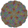
Journal: Nat Commun / Year: 2025 Title: Structural insights into scaffold-guided assembly of the Pseudomonas phage D3 capsid. Authors: Anna K Belford / Joshua B Maurer / Robert L Duda / Alexis Huet / James F Conway / Abstract: Tailed bacteriophages comprise the largest structural family of viruses with close relatives in archaea and the eukaryotic herpesviruses. The common assembly pathway produces an icosahedrally ...Tailed bacteriophages comprise the largest structural family of viruses with close relatives in archaea and the eukaryotic herpesviruses. The common assembly pathway produces an icosahedrally symmetric protein shell, called capsid, into which the double-stranded DNA genome is packaged. While capsid sizes and amino acid sequences vary considerably, the major capsid protein (MCP) folds are remarkably similar throughout the family. To investigate the mechanisms governing capsid size, we characterize the procapsid and mature capsid of phage D3, which expresses an icosahedral lattice with Triangulation number T = 9. We find that the MCP scaffold domain binds to the interior capsid surface, acting as a clamp to constrain subunit interactions. Following scaffold digestion, the MCP capsid domains form strong interactions that maintain capsid structure throughout maturation. The scaffold constraints appear critical for capsid size determination and provide important understanding of the factors governing capsid assembly in general and expands our understanding of these ecologically and biomedically important viruses.
History
Deposition
May 29, 2025
Deposition site: RCSB / Processing site: RCSB
Revision 1.0
Mar 11, 2026
Provider: repository / Type: Initial release
Revision 1.0
Mar 11, 2026
Data content type: EM metadata / Data content type: EM metadata / Provider: repository / Type: Initial release
Revision 1.0
Mar 11, 2026
Data content type: FSC / Data content type: FSC / Provider: repository / Type: Initial release
Revision 1.0
Mar 11, 2026
Data content type: Half map / Part number: 1 / Data content type: Half map / Provider: repository / Type: Initial release
Revision 1.0
Mar 11, 2026
Data content type: Half map / Part number: 2 / Data content type: Half map / Provider: repository / Type: Initial release
Revision 1.0
Mar 11, 2026
Data content type: Image / Data content type: Image / Provider: repository / Type: Initial release
Revision 1.0
Mar 11, 2026
Data content type: Primary map / Data content type: Primary map / Provider: repository / Type: Initial release
#200 - Aug 2016 Quasisymmetry in Icosahedral Viruses similarity (2)
-
Assembly
Deposited unit
A: Major capsid protein B: Major capsid protein C: Major capsid protein D: Major capsid protein E: Major capsid protein F: Major capsid protein G: Major capsid protein H: Major capsid protein I: Major capsid protein
A: Major capsid protein B: Major capsid protein C: Major capsid protein D: Major capsid protein E: Major capsid protein F: Major capsid protein G: Major capsid protein H: Major capsid protein I: Major capsid protein
A: Major capsid protein B: Major capsid protein C: Major capsid protein D: Major capsid protein E: Major capsid protein F: Major capsid protein G: Major capsid protein H: Major capsid protein I: Major capsid protein
x 5
icosahedral pentamer
1.93 MDa, 45 polymers
Theoretical mass
Number of molelcules
Total (without water)
1,929,237
45
Polymers
1,929,237
45
Non-polymers
0
0
Water
0
Type
Name
Symmetry operation
Number
identity operation
1_555
x,y,z
1
point symmetry operation
4
4
A: Major capsid protein B: Major capsid protein C: Major capsid protein D: Major capsid protein E: Major capsid protein F: Major capsid protein G: Major capsid protein H: Major capsid protein I: Major capsid protein
x 6
icosahedral 23 hexamer
2.32 MDa, 54 polymers
Theoretical mass
Number of molelcules
Total (without water)
2,315,084
54
Polymers
2,315,084
54
Non-polymers
0
0
Water
0
Type
Name
Symmetry operation
Number
identity operation
1_555
x,y,z
1
point symmetry operation
5
5
Idetical with deposited unit in distinct coordinate
icosahedral asymmetric unit, std point frame
Type
Name
Symmetry operation
Number
transform to point frame
1
Symmetry
Point symmetry: (Schoenflies symbol: I (icosahedral))
-
Components
#1: Protein
Majorcapsidprotein
Mass: 42871.926 Da / Num. of mol.: 9 / Source method: isolated from a natural source / Source: (natural) Pseudomonas phage D3 (virus) / References: UniProt: Q9XJT3
Has protein modification
N
-
Experimental details
-
Experiment
Experiment
Method: ELECTRON MICROSCOPY
EM experiment
Aggregation state: PARTICLE / 3D reconstruction method: single particle reconstruction
Electron dose: 60 e/Å2 / Detector mode: COUNTING / Film or detector model: FEI FALCON IV (4k x 4k) / Num. of grids imaged: 1 / Num. of real images: 43546
-
Processing
EM software
ID
Name
Version
Category
1
EMAN2
particleselection
2
EPU
imageacquisition
4
CTFFIND
CTFcorrection
7
UCSF ChimeraX
1.9
modelfitting
9
RELION
4
initialEulerassignment
10
RELION
4
finalEulerassignment
12
RELION
4
3Dreconstruction
13
ISOLDE
modelrefinement
CTF correction
Type: PHASE FLIPPING AND AMPLITUDE CORRECTION
Symmetry
Point symmetry: I (icosahedral)
3D reconstruction
Resolution: 3.5 Å / Resolution method: FSC 0.143 CUT-OFF / Num. of particles: 1584 / Symmetry type: POINT
In the structure databanks used in Yorodumi, some data are registered as the other names, "COVID-19 virus" and "2019-nCoV". Here are the details of the virus and the list of structure data.
Jan 31, 2019. EMDB accession codes are about to change! (news from PDBe EMDB page)
EMDB accession codes are about to change! (news from PDBe EMDB page)
The allocation of 4 digits for EMDB accession codes will soon come to an end. Whilst these codes will remain in use, new EMDB accession codes will include an additional digit and will expand incrementally as the available range of codes is exhausted. The current 4-digit format prefixed with “EMD-” (i.e. EMD-XXXX) will advance to a 5-digit format (i.e. EMD-XXXXX), and so on. It is currently estimated that the 4-digit codes will be depleted around Spring 2019, at which point the 5-digit format will come into force.
The EM Navigator/Yorodumi systems omit the EMD- prefix.
Related info.:Q: What is EMD? / ID/Accession-code notation in Yorodumi/EM Navigator
Yorodumi is a browser for structure data from EMDB, PDB, SASBDB, etc.
This page is also the successor to EM Navigator detail page, and also detail information page/front-end page for Omokage search.
The word "yorodu" (or yorozu) is an old Japanese word meaning "ten thousand". "mi" (miru) is to see.
Related info.:EMDB / PDB / SASBDB / Comparison of 3 databanks / Yorodumi Search / Aug 31, 2016. New EM Navigator & Yorodumi / Yorodumi Papers / Jmol/JSmol / Function and homology information / Changes in new EM Navigator and Yorodumi
 Movie
Movie Controller


 Open data
 Basic information
 Components
 Keywords
 Function and homology information
 Pseudomonas phage D3 (virus)
 Authors
 United States, 2items
  Citation
 Structure visualization
 Downloads & links
 Other downloads


 PDBj
PDBj

 Assembly


 Sample preparation
 Electron microscopy imaging
 FIELD EMISSION GUN / Accelerating voltage: 300 kV / Illumination mode: FLOOD BEAM
 Processing