Movie
Movie Controller


+ Open data

- Basic information
| Entry | Database: PDB / ID: 9orz | ||||||||||||
|---|---|---|---|---|---|---|---|---|---|---|---|---|---|
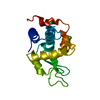
| Title | MicroED structure of apo-form lysozyme | ||||||||||||
 Components Components | Lysozyme C | ||||||||||||
 Keywords Keywords | HYDROLASE / Enzyme / MicroED | ||||||||||||
| Function / homology |  Function and homology information Function and homology informationLactose synthesis / Antimicrobial peptides / Neutrophil degranulation / beta-N-acetylglucosaminidase activity / cell wall macromolecule catabolic process / lysozyme / lysozyme activity / killing of cells of another organism / defense response to Gram-negative bacterium / defense response to bacterium ...Lactose synthesis / Antimicrobial peptides / Neutrophil degranulation / beta-N-acetylglucosaminidase activity / cell wall macromolecule catabolic process / lysozyme / lysozyme activity / killing of cells of another organism / defense response to Gram-negative bacterium / defense response to bacterium / defense response to Gram-positive bacterium / endoplasmic reticulum / : / identical protein binding / cytoplasm Similarity search - Function | ||||||||||||
| Biological species |  | ||||||||||||
| Method | ELECTRON CRYSTALLOGRAPHY / electron crystallography /  MOLECULAR REPLACEMENT / cryo EM / Resolution: 2.3 Å MOLECULAR REPLACEMENT / cryo EM / Resolution: 2.3 Å | ||||||||||||
 Authors Authors | Vlahakis, N.W. / Flowers, C.W. / Rodriguez, J.A. | ||||||||||||
| Funding support |  United States, 3items United States, 3items
| ||||||||||||
 Citation Citation | Journal: Proc Natl Acad Sci U S A / Year: 2025 Title: Combining MicroED and native mass spectrometry for structural discovery of enzyme-small molecule complexes. Authors: Niko W Vlahakis / Cameron W Flowers / Mengting Liu / Matthew P Agdanowski / Samuel Johnson / Jacob A Summers / Lian M C Jacobs / Catherine Keyser / Phoebe Russell / Samuel L Rose / Julien ...Authors: Niko W Vlahakis / Cameron W Flowers / Mengting Liu / Matthew P Agdanowski / Samuel Johnson / Jacob A Summers / Lian M C Jacobs / Catherine Keyser / Phoebe Russell / Samuel L Rose / Julien Orlans / Nima Adhami / Yu Chen / Michael R Sawaya / Shibom Basu / Daniele de Sanctis / Yu Chen / Soichi Wakatsuki / Hosea M Nelson / Joseph A Loo / Yi Tang / Jose A Rodriguez /  Abstract: With the goal of accelerating the discovery of small molecule-protein complexes, we leverage fast, low-dose, event-based electron counting microcrystal electron diffraction (MicroED) data collection ...With the goal of accelerating the discovery of small molecule-protein complexes, we leverage fast, low-dose, event-based electron counting microcrystal electron diffraction (MicroED) data collection and native mass spectrometry. This approach, which we term electron diffraction with native mass spectrometry (ED-MS), allows assignment of protein target structures bound to ligands with data obtained from crystal slurries soaked with mixtures of known inhibitors and crude biosynthetic reactions. This extends to libraries of printed ligands dispensed directly onto TEM grids for later soaking with microcrystal slurries, and complexes with noncovalent ligands. ED-MS resolves structures of the natural product, epoxide-based cysteine protease inhibitor E-64, and its biosynthetic analogs bound to the model cysteine protease, papain. It further identifies papain binding to its preferred natural products, by showing that two analogs of E-64 outcompete others in binding to papain crystals, and by detecting papain bound to E-64 and an analog from crude biosynthetic reactions, without purification. ED-MS also resolves binding of the CTX-M-14 β-lactamase, a target of active drug development, to the non-β-lactam inhibitor, avibactam, alone or in a cocktail of unrelated compounds. These results illustrate the utility of ED-MS for natural product ligand discovery and for structure-based screening of small molecule binders to macromolecular targets, promising utility for drug discovery. | ||||||||||||
| History |
|
- Structure visualization
| Structure viewer | Molecule: MolmilJmol/JSmol |
|---|
- Downloads & links
-Download
| PDBx/mmCIF format | 9orz.cif.gz | 39.6 KB | Display | PDBx/mmCIF format |
|---|---|---|---|---|
| PDB format | pdb9orz.ent.gz | 24.4 KB | Display | PDB format |
| PDBx/mmJSON format | 9orz.json.gz | Tree view | PDBx/mmJSON format | |
| Others |  Other downloads Other downloads |
-Validation report
| Arichive directory | https://data.pdbj.org/pub/pdb/validation_reports/or/9orzftp://data.pdbj.org/pub/pdb/validation_reports/or/9orz | HTTPS FTP |
|---|
-Related structure data
| Related structure data |  9nbpC  9nbqC  9nc1C  9ncaC  9nccC  9oqeC  9or3C  9or7C  9orbC  9orgC  9orhC  9orlC  9orsC  9orvC  9orwC  9orxC  9oryC  9os0C  9os1C  9os8C C: citing same article ( |
|---|---|
| Similar structure data |
-Links
 PDBj
PDBj


- Assembly
| Deposited unit | 
| ||||||||
|---|---|---|---|---|---|---|---|---|---|
| 1 |
| ||||||||
| Unit cell |
|
-Components
| #1: Protein | Mass: 14331.160 Da / Num. of mol.: 1 / Fragment: lyzozyme / Source method: isolated from a natural source / Source: (natural) |
|---|---|
| #2: Chemical | ChemComp-NA /   Mass: 22.990 Da / Num. of mol.: 1 / Source method: obtained synthetically / Formula: Na Mass: 22.990 Da / Num. of mol.: 1 / Source method: obtained synthetically / Formula: Na |
| #3: Water | ChemComp-HOH /  Mass: 18.015 Da / Num. of mol.: 15 / Source method: isolated from a natural source / Formula: H2O Mass: 18.015 Da / Num. of mol.: 15 / Source method: isolated from a natural source / Formula: H2O |
| Has ligand of interest | N |
| Has protein modification | Y |
-Experimental details
-Experiment
| Experiment | Method: ELECTRON CRYSTALLOGRAPHY |
|---|---|
| EM experiment | Aggregation state: 3D ARRAY / 3D reconstruction method: electron crystallography |
- Sample preparation
| Component | Name: Lysozyme / Type: COMPLEX Details: Crystals formed by vapor diffusion in sitting drops, from 80 mg/mL lysozyme mixed 1:1 with reservoir solution (800 mM NaCl, 80 mM sodium acetate pH 4.8) Entity ID: #1 / Source: NATURAL |
|---|---|
| Source (natural) | Organism: |
| EM crystal formation | Details: Crystals formed by vapor diffusion in sitting drops, from 80 mg/mL lysozyme mixed 1:1 with reservoir solution (800 mM NaCl, 80 mM sodium acetate pH 4.8) |
| Buffer solution | pH: 4.8 / Details: 800 mM NaCl, 80 mM sodium acetate pH 4.8 |
| Specimen | Embedding applied: NO / Shadowing applied: NO / Staining applied: NO / Vitrification applied: YES |
| Vitrification | Cryogen name: ETHANE |
-Data collection
| Experimental equipment |  Model: Tecnai F20 / Image courtesy: FEI Company |
|---|---|
| Microscopy | Model: TFS TALOS F200C |
| Electron gun | Electron source: FIELD EMISSION GUN / Accelerating voltage: 200 kV / Illumination mode: FLOOD BEAM |
| Electron lens | Mode: DIFFRACTION / Nominal defocus max: 0 nm / Nominal defocus min: 0 nm / C2 aperture diameter: 70 µm |
| Image recording | Electron dose: 0.09 e/Å2 / Film or detector model: DIRECT ELECTRON APOLLO (4k x 4k) |
| EM diffraction shell | Resolution: 2.3→2.4 Å / Fourier space coverage: 95.6 % / Multiplicity: 6 / Num. of structure factors: 603 / Phase residual: 30.8 ° |
| EM diffraction stats | Fourier space coverage: 92.9 % / High resolution: 2.3 Å / Num. of intensities measured: 30058 / Num. of structure factors: 5060 / Phase error rejection criteria: NONE / Rmerge: 0.252 |
- Processing
| Software |
| |||||||||||||||||||||||||||||||||||
|---|---|---|---|---|---|---|---|---|---|---|---|---|---|---|---|---|---|---|---|---|---|---|---|---|---|---|---|---|---|---|---|---|---|---|---|---|
| EM 3D crystal entity | ∠α: 90 ° / ∠β: 90 ° / ∠γ: 90 ° / A: 77.53 Å / B: 77.53 Å / C: 37.33 Å / Space group name: P43212 / Space group num: 96 | |||||||||||||||||||||||||||||||||||
| CTF correction | Type: NONE | |||||||||||||||||||||||||||||||||||
| 3D reconstruction | Resolution: 2.3 Å / Resolution method: DIFFRACTION PATTERN/LAYERLINES / Symmetry type: 3D CRYSTAL | |||||||||||||||||||||||||||||||||||
| Atomic model building | PDB-ID: 1DPX Accession code: 1DPX / Source name: PDB / Type: experimental model | |||||||||||||||||||||||||||||||||||
| Refinement | Method to determine structure: MOLECULAR REPLACEMENT / Resolution: 2.3→54.82 Å / SU ML: 0.28 / Cross valid method: FREE R-VALUE / σ(F): 1.36 / Phase error: 24.87 / Stereochemistry target values: ML
| |||||||||||||||||||||||||||||||||||
| Solvent computation | Shrinkage radii: 0.9 Å / VDW probe radii: 1.1 Å / Solvent model: FLAT BULK SOLVENT MODEL | |||||||||||||||||||||||||||||||||||
| Refine LS restraints |
| |||||||||||||||||||||||||||||||||||
| LS refinement shell |
|