 Movie
Movie Controller


+ Open data

- Basic information
| Entry | Database: PDB / ID: 9olz | ||||||
|---|---|---|---|---|---|---|---|
| Title | Crystal structure of E. coli ApaH in complex with Ap4A | ||||||
 Components Components | Bis(5'-nucleosyl)-tetraphosphatase [symmetrical] | ||||||
 Keywords Keywords | HYDROLASE / ApaH / symmetrical hydrolase / RNA decapping | ||||||
| Function / homology |  Function and homology information Function and homology informationbis(5'-nucleosyl)-tetraphosphatase activity / bis(5'-nucleosyl)-tetraphosphatase (symmetrical) / bis(5'-nucleosyl)-tetraphosphatase (symmetrical) activity / RNA decapping / nucleobase-containing small molecule interconversion / phosphatase activity / response to X-ray / hydrolase activity / cytoplasm Similarity search - Function | ||||||
| Biological species |  | ||||||
| Method |  X-RAY DIFFRACTION / SYNCHROTRON / MOLECULAR REPLACEMENT / Resolution: 1.71 Å X-RAY DIFFRACTION / SYNCHROTRON / MOLECULAR REPLACEMENT / Resolution: 1.71 Å | ||||||
 Authors Authors | Nuthanakanti, A. / Serganov, A. | ||||||
| Funding support |  United States, 1items United States, 1items
| ||||||
 Citation Citation | Journal: Nat.Chem.Biol. / Year: 2026 Title: ApaH decaps Np 4 N-capped RNAs in two alternative orientations. Authors: Nuthanakanti, A. / Korn, M. / Levenson-Palmer, R. / Wu, Y. / Babu, N.R. / Huang, X. / Banh, R.S. / Belasco, J.G. / Serganov, A. | ||||||
| History |
|
- Structure visualization
| Structure viewer | Molecule: MolmilJmol/JSmol |
|---|
- Downloads & links
-Download
| PDBx/mmCIF format | 9olz.cif.gz | 166.2 KB | Display | PDBx/mmCIF format |
|---|---|---|---|---|
| PDB format | pdb9olz.ent.gz | 103.7 KB | Display | PDB format |
| PDBx/mmJSON format | 9olz.json.gz | Tree view | PDBx/mmJSON format | |
| Others |  Other downloads Other downloads |
-Validation report
| Arichive directory | https://data.pdbj.org/pub/pdb/validation_reports/ol/9olzftp://data.pdbj.org/pub/pdb/validation_reports/ol/9olz | HTTPS FTP |
|---|
-Related structure data
| Related structure data |  9ojdC  9ojpC  9ojqC  9ojwC  9ojxC  9ok1C  9ok2C  9olnC  9olyC  9om9C  9omcC  9omuC  9omwC  9omxC  9on0C  9on7C  9ondC  9ongC  9oonC  9ooyC  9op2C  9opgC  9ophC  9oq9C  9oqbC C: citing same article ( |
|---|---|
| Similar structure data |
-Links
 PDBj
PDBj
- Assembly
| Deposited unit | 
| |||||||||||||||||||||||||||||||||||||||||||||||||||||||||||||||||||||||||||||||
|---|---|---|---|---|---|---|---|---|---|---|---|---|---|---|---|---|---|---|---|---|---|---|---|---|---|---|---|---|---|---|---|---|---|---|---|---|---|---|---|---|---|---|---|---|---|---|---|---|---|---|---|---|---|---|---|---|---|---|---|---|---|---|---|---|---|---|---|---|---|---|---|---|---|---|---|---|---|---|---|---|
| 1 | 
| |||||||||||||||||||||||||||||||||||||||||||||||||||||||||||||||||||||||||||||||
| 2 | 
| |||||||||||||||||||||||||||||||||||||||||||||||||||||||||||||||||||||||||||||||
| Unit cell |
| |||||||||||||||||||||||||||||||||||||||||||||||||||||||||||||||||||||||||||||||
| Noncrystallographic symmetry (NCS) | NCS domain:
NCS domain segments: Ens-ID: ens_1
NCS oper: (Code: givenMatrix: (-0.0281364993886, -0.0506135166852, 0.99832189665), (0.0323857930984, -0.998239155516, -0.0496965672818), (0.999079325083, 0.0309331589556, 0.0297261142682)Vector: 3. ...NCS oper: (Code: given Matrix: (-0.0281364993886, -0.0506135166852, 0.99832189665), Vector: |
-Components
-Protein , 1 types, 2 molecules AB
| #1: Protein | Mass: 31331.666 Da / Num. of mol.: 2 Source method: isolated from a genetically manipulated source Source: (gene. exp.) References: UniProt: P05637, bis(5'-nucleosyl)-tetraphosphatase (symmetrical) |
|---|
-Non-polymers , 5 types, 478 molecules 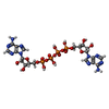


| #2: Chemical |  Mass: 836.387 Da / Num. of mol.: 2 / Source method: obtained synthetically / Formula: C20H28N10O19P4 / Feature type: SUBJECT OF INVESTIGATION Mass: 836.387 Da / Num. of mol.: 2 / Source method: obtained synthetically / Formula: C20H28N10O19P4 / Feature type: SUBJECT OF INVESTIGATION#3: Chemical | ChemComp-MN /  Mass: 54.938 Da / Num. of mol.: 4 / Source method: obtained synthetically / Formula: Mn Mass: 54.938 Da / Num. of mol.: 4 / Source method: obtained synthetically / Formula: Mn#4: Chemical |  Mass: 24.305 Da / Num. of mol.: 2 / Source method: obtained synthetically / Formula: Mg Mass: 24.305 Da / Num. of mol.: 2 / Source method: obtained synthetically / Formula: Mg#5: Chemical | ChemComp-EPE / |  Mass: 238.305 Da / Num. of mol.: 1 / Source method: obtained synthetically / Formula: C8H18N2O4S / Comment: pH buffer*YM Mass: 238.305 Da / Num. of mol.: 1 / Source method: obtained synthetically / Formula: C8H18N2O4S / Comment: pH buffer*YM#6: Water | ChemComp-HOH / | Mass: 18.015 Da / Num. of mol.: 469 / Source method: isolated from a natural source / Formula: H2O |
|---|
-Details
| Has ligand of interest | Y |
|---|---|
| Has protein modification | N |
-Experimental details
-Experiment
| Experiment | Method: X-RAY DIFFRACTION / Number of used crystals: 1 |
|---|
- Sample preparation
| Crystal | Density Matthews: 3.44 Å3/Da / Density % sol: 64.21 % |
|---|---|
| Crystal grow | Temperature: 291 K / Method: vapor diffusion, hanging drop / pH: 7.5 Details: Conditions: 0.3 mM ApaH (10 mg/mL), 1 mM DTT, 5 mM MgCl2, 25 mM Hepes (pH 7.5), 0.2 M NaCl and soaked with Ap4A Well solution: 0.2 M Sodium malonate, 20% PEG3350. |
-Data collection
| Diffraction | Mean temperature: 100 K / Serial crystal experiment: N |
|---|---|
| Diffraction source | Source: SYNCHROTRON / Site: APS / Beamline: 24-ID-C / Wavelength: 0.9791 Å |
| Detector | Type: DECTRIS EIGER2 X 16M / Detector: PIXEL / Date: Jul 18, 2019 |
| Radiation | Protocol: SINGLE WAVELENGTH / Monochromatic (M) / Laue (L): M / Scattering type: x-ray |
| Radiation wavelength | Wavelength: 0.9791 Å / Relative weight: 1 |
| Reflection | Resolution: 1.71→83.67 Å / Num. obs: 91253 / % possible obs: 98.3 % / Redundancy: 5 % / Biso Wilson estimate: 31.6 Å2 / CC1/2: 0.995 / Rmerge(I) obs: 0.08 / Net I/σ(I): 10.4 |
| Reflection shell | Resolution: 1.71→1.73 Å / Redundancy: 4.9 % / Rmerge(I) obs: 1.739 / Mean I/σ(I) obs: 0.6 / Num. unique obs: 4085 / CC1/2: 0.253 / % possible all: 89.1 |
- Processing
| Software |
| ||||||||||||||||||||||||||||||||||||||||||||||||||||||||||||||||||||||||||||||||||||||||||||||||||||||||||||||||||||||||||||||||||||||||||||||||||||||||||||||||||||||||||||||||||||||||||||||||||||
|---|---|---|---|---|---|---|---|---|---|---|---|---|---|---|---|---|---|---|---|---|---|---|---|---|---|---|---|---|---|---|---|---|---|---|---|---|---|---|---|---|---|---|---|---|---|---|---|---|---|---|---|---|---|---|---|---|---|---|---|---|---|---|---|---|---|---|---|---|---|---|---|---|---|---|---|---|---|---|---|---|---|---|---|---|---|---|---|---|---|---|---|---|---|---|---|---|---|---|---|---|---|---|---|---|---|---|---|---|---|---|---|---|---|---|---|---|---|---|---|---|---|---|---|---|---|---|---|---|---|---|---|---|---|---|---|---|---|---|---|---|---|---|---|---|---|---|---|---|---|---|---|---|---|---|---|---|---|---|---|---|---|---|---|---|---|---|---|---|---|---|---|---|---|---|---|---|---|---|---|---|---|---|---|---|---|---|---|---|---|---|---|---|---|---|---|---|---|
| Refinement | Method to determine structure: MOLECULAR REPLACEMENT / Resolution: 1.71→83.67 Å / SU ML: 0.2797 / Cross valid method: FREE R-VALUE / σ(F): 1.34 / Phase error: 26.1059 Stereochemistry target values: GeoStd + Monomer Library + CDL v1.2
| ||||||||||||||||||||||||||||||||||||||||||||||||||||||||||||||||||||||||||||||||||||||||||||||||||||||||||||||||||||||||||||||||||||||||||||||||||||||||||||||||||||||||||||||||||||||||||||||||||||
| Solvent computation | Shrinkage radii: 0.9 Å / VDW probe radii: 1.11 Å / Solvent model: FLAT BULK SOLVENT MODEL | ||||||||||||||||||||||||||||||||||||||||||||||||||||||||||||||||||||||||||||||||||||||||||||||||||||||||||||||||||||||||||||||||||||||||||||||||||||||||||||||||||||||||||||||||||||||||||||||||||||
| Displacement parameters | Biso mean: 35.89 Å2 | ||||||||||||||||||||||||||||||||||||||||||||||||||||||||||||||||||||||||||||||||||||||||||||||||||||||||||||||||||||||||||||||||||||||||||||||||||||||||||||||||||||||||||||||||||||||||||||||||||||
| Refinement step | Cycle: LAST / Resolution: 1.71→83.67 Å
| ||||||||||||||||||||||||||||||||||||||||||||||||||||||||||||||||||||||||||||||||||||||||||||||||||||||||||||||||||||||||||||||||||||||||||||||||||||||||||||||||||||||||||||||||||||||||||||||||||||
| Refine LS restraints |
| ||||||||||||||||||||||||||||||||||||||||||||||||||||||||||||||||||||||||||||||||||||||||||||||||||||||||||||||||||||||||||||||||||||||||||||||||||||||||||||||||||||||||||||||||||||||||||||||||||||
| Refine LS restraints NCS | Type: Torsion NCS / Rms dev position: 0.576875335739 Å | ||||||||||||||||||||||||||||||||||||||||||||||||||||||||||||||||||||||||||||||||||||||||||||||||||||||||||||||||||||||||||||||||||||||||||||||||||||||||||||||||||||||||||||||||||||||||||||||||||||
| LS refinement shell |
|
