+ Open data
- Basic information
| Entry | Database: PDB / ID: 9oh5 | ||||||||||||||||||||||||
|---|---|---|---|---|---|---|---|---|---|---|---|---|---|---|---|---|---|---|---|---|---|---|---|---|---|
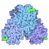
| Title | Cryo-EM structure of the assembled MS2 CPM58 VLP | ||||||||||||||||||||||||
 Components Components | MS2 CPM58 | ||||||||||||||||||||||||
 Keywords Keywords | VIRUS LIKE PARTICLE / bacteriophage / circular permutation | ||||||||||||||||||||||||
| Function / homology |  Function and homology information Function and homology informationnegative regulation of viral translation / T=3 icosahedral viral capsid / regulation of translation / structural molecule activity / RNA binding / identical protein binding Similarity search - Function | ||||||||||||||||||||||||
| Biological species |  Escherichia phage MS2 (virus) Escherichia phage MS2 (virus) | ||||||||||||||||||||||||
| Method | ELECTRON MICROSCOPY / single particle reconstruction / cryo EM / Resolution: 2.62 Å | ||||||||||||||||||||||||
 Authors Authors | Liang, S. / Jung, J. / Tullman-Ercek, D. | ||||||||||||||||||||||||
| Funding support |  United States, 1items United States, 1items
| ||||||||||||||||||||||||
 Citation Citation |  Journal: ACS Nano / Year: 2025 Journal: ACS Nano / Year: 2025Title: Synthetic Rewiring of Virus-like Particles via Circular Permutation Enables Modular Peptide Display and Protein Encapsulation. Authors: Shiqi Liang / Kaavya Butaney / Daniel de Castro Assumpção / James Jung / Nolan W Kennedy / Danielle Tullman-Ercek /  Abstract: Virus-like particles (VLPs) are self-assembling nanoparticles derived from viruses with the potential as scaffolds for myriad applications. They are also excellent testbeds for engineering protein ...Virus-like particles (VLPs) are self-assembling nanoparticles derived from viruses with the potential as scaffolds for myriad applications. They are also excellent testbeds for engineering protein superstructures. Engineers often employ techniques such as amino acid substitutions and insertions/deletions. Yet evolution also utilizes circular permutation, a powerful natural strategy that has not been fully explored in engineering self-assembling protein nanoparticles. Here, we demonstrate this technique using the MS2 VLP as a model self-assembling proteinaceous nanoparticle. We constructed a comprehensive circular permutation library of the fused MS2 coat protein dimer construct. The strategy revealed terminal locations, validated via cryo-electron microscopy, that enabled C-terminal peptide tagging and led to a protein encapsulation strategy via covalent bonding - a feature the native coat protein does not permit. Our systematic study demonstrates the power of circular permutation for engineering features as well as quantitatively and systematically exploring VLP structural determinants. | ||||||||||||||||||||||||
| History |
|
- Structure visualization
| Structure viewer | Molecule:  Molmil Molmil Jmol/JSmol Jmol/JSmol |
|---|
- Downloads & links
- Download
| PDBx/mmCIF format |  9oh5.cif.gz 9oh5.cif.gz | 85.5 KB | Display |  PDBx/mmCIF format PDBx/mmCIF format |
|---|---|---|---|---|
| PDB format |  pdb9oh5.ent.gz pdb9oh5.ent.gz | 62.7 KB | Display |  PDB format PDB format |
| PDBx/mmJSON format |  9oh5.json.gz 9oh5.json.gz | Tree view |  PDBx/mmJSON format PDBx/mmJSON format | |
| Others |  Other downloads Other downloads |
-Validation report
| Summary document |  9oh5_validation.pdf.gz 9oh5_validation.pdf.gz | 1.3 MB | Display |  wwPDB validaton report wwPDB validaton report |
|---|---|---|---|---|
| Full document |  9oh5_full_validation.pdf.gz 9oh5_full_validation.pdf.gz | 1.3 MB | Display | |
| Data in XML |  9oh5_validation.xml.gz 9oh5_validation.xml.gz | 36.8 KB | Display | |
| Data in CIF |  9oh5_validation.cif.gz 9oh5_validation.cif.gz | 51.5 KB | Display | |
| Arichive directory |  https://data.pdbj.org/pub/pdb/validation_reports/oh/9oh5 https://data.pdbj.org/pub/pdb/validation_reports/oh/9oh5 ftp://data.pdbj.org/pub/pdb/validation_reports/oh/9oh5 ftp://data.pdbj.org/pub/pdb/validation_reports/oh/9oh5 | HTTPS FTP |
-Related structure data
| Related structure data |  70484MC M: map data used to model this data C: citing same article ( |
|---|---|
| Similar structure data | Similarity search - Function & homology  F&H Search F&H Search |
- Links
Links
- Assembly
| Deposited unit | 
|
|---|---|
| 1 | x 60
|
| 2 |
|
| 3 | x 5
|
| 4 | x 6
|
| 5 | 
|
| Symmetry | Point symmetry: (Schoenflies symbol: I (icosahedral)) |
- Components
| #1: Protein | Mass: 27458.957 Da / Num. of mol.: 2 Source method: isolated from a genetically manipulated source Source: (gene. exp.)  Escherichia phage MS2 (virus) / Production host: Escherichia phage MS2 (virus) / Production host:  Has protein modification | N | |
|---|
-Experimental details
-Experiment
| Experiment | Method: ELECTRON MICROSCOPY |
|---|---|
| EM experiment | Aggregation state: PARTICLE / 3D reconstruction method: single particle reconstruction |
- Sample preparation
| Component | Name: Escherichia phage MS2 / Type: VIRUS / Entity ID: all / Source: RECOMBINANT |
|---|---|
| Molecular weight | Value: 2.4696 MDa / Experimental value: NO |
| Source (natural) | Organism:  Escherichia phage MS2 (virus) Escherichia phage MS2 (virus) |
| Source (recombinant) | Organism:  |
| Details of virus | Empty: YES / Enveloped: NO / Isolate: STRAIN / Type: VIRUS-LIKE PARTICLE |
| Virus shell | Name: CPM58 VLP / Diameter: 300 nm / Triangulation number (T number): 3 |
| Buffer solution | pH: 7.2 |
| Specimen | Conc.: 2 mg/ml / Embedding applied: NO / Shadowing applied: NO / Staining applied: NO / Vitrification applied: YES |
| Specimen support | Grid material: COPPER / Grid mesh size: 400 divisions/in. / Grid type: Quantifoil R1.2/1.3 |
| Vitrification | Instrument: FEI VITROBOT MARK IV / Cryogen name: ETHANE / Humidity: 95 % / Chamber temperature: 288 K |
- Electron microscopy imaging
| Microscopy | Model: TFS GLACIOS |
|---|---|
| Electron gun | Electron source:  FIELD EMISSION GUN / Accelerating voltage: 200 kV / Illumination mode: FLOOD BEAM FIELD EMISSION GUN / Accelerating voltage: 200 kV / Illumination mode: FLOOD BEAM |
| Electron lens | Mode: BRIGHT FIELD / Nominal magnification: 130000 X / Nominal defocus max: 1600 nm / Nominal defocus min: 600 nm / Cs: 2.7 mm / C2 aperture diameter: 20 µm / Alignment procedure: COMA FREE |
| Specimen holder | Cryogen: NITROGEN / Specimen holder model: FEI TITAN KRIOS AUTOGRID HOLDER / Temperature (min): 77 K |
| Image recording | Average exposure time: 5.33 sec. / Electron dose: 60 e/Å2 / Film or detector model: TFS FALCON 4i (4k x 4k) / Num. of grids imaged: 1 / Num. of real images: 5849 |
| EM imaging optics | Energyfilter name: TFS Selectris / Energyfilter slit width: 10 eV |
- Processing
| EM software |
| ||||||||||||||||||||||||||||||||||||||||
|---|---|---|---|---|---|---|---|---|---|---|---|---|---|---|---|---|---|---|---|---|---|---|---|---|---|---|---|---|---|---|---|---|---|---|---|---|---|---|---|---|---|
| CTF correction | Type: PHASE FLIPPING AND AMPLITUDE CORRECTION | ||||||||||||||||||||||||||||||||||||||||
| Particle selection | Num. of particles selected: 539975 | ||||||||||||||||||||||||||||||||||||||||
| Symmetry | Point symmetry: I (icosahedral) | ||||||||||||||||||||||||||||||||||||||||
| 3D reconstruction | Resolution: 2.62 Å / Resolution method: FSC 0.143 CUT-OFF / Num. of particles: 109941 / Algorithm: BACK PROJECTION / Num. of class averages: 1 / Symmetry type: POINT | ||||||||||||||||||||||||||||||||||||||||
| Atomic model building | Protocol: FLEXIBLE FIT / Space: REAL | ||||||||||||||||||||||||||||||||||||||||
| Atomic model building | PDB-ID: 1ZDI Accession code: 1ZDI / Source name: PDB / Type: experimental model | ||||||||||||||||||||||||||||||||||||||||
| Refinement | Highest resolution: 2.62 Å Stereochemistry target values: REAL-SPACE (WEIGHTED MAP SUM AT ATOM CENTERS) | ||||||||||||||||||||||||||||||||||||||||
| Refine LS restraints |
|
 Movie
Movie Controller


 PDBj
PDBj


