 Movie
Movie Controller

[English] 日本語
 Yorodumi
- PDB-9of6: Structure of the Acinetobacter baumannii Response Regulator PmrA ... -
+ Open data

- Basic information
| Entry | Database: PDB / ID: 9of6 | ||||||
|---|---|---|---|---|---|---|---|
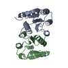
| Title | Structure of the Acinetobacter baumannii Response Regulator PmrA Receiver Domain I13M Mutation in Active Dimer State | ||||||
 Components Components | PmrA | ||||||
 Keywords Keywords | TRANSCRIPTION / Response regulator / two component system / Polymyxin resistance / Resistant mutant | ||||||
| Function / homology |  Function and homology information Function and homology informationphosphorelay response regulator activity / protein-DNA complex / transcription cis-regulatory region binding / regulation of DNA-templated transcription / cytosol Similarity search - Function | ||||||
| Biological species |  Acinetobacter baumannii (bacteria) Acinetobacter baumannii (bacteria) | ||||||
| Method |  X-RAY DIFFRACTION / SYNCHROTRON / MOLECULAR REPLACEMENT / Resolution: 1.86 Å X-RAY DIFFRACTION / SYNCHROTRON / MOLECULAR REPLACEMENT / Resolution: 1.86 Å | ||||||
 Authors Authors | Milton, M.E. / Jaimes, F.E. / Cavanagh, J. | ||||||
| Funding support |  United States, 1items United States, 1items
| ||||||
 Citation Citation | Journal: Microorganisms / Year: 2025 Title: PmrA Mutations in Drug-Resistant Acinetobacter baumannii Affect Sensor Kinase-Response Regulator Interaction and Phosphotransfer Authors: Jaimes, F.E. / Hondros, A.D. / Kinkead, J. / Milton, M.E. / Thompson, R.J. / Figg, A.M. / Melander, C. / Cavanagh, J. | ||||||
| History |
|
- Structure visualization
| Structure viewer | Molecule: MolmilJmol/JSmol |
|---|
- Downloads & links
-Download
| PDBx/mmCIF format | 9of6.cif.gz | 130 KB | Display | PDBx/mmCIF format |
|---|---|---|---|---|
| PDB format | pdb9of6.ent.gz | 82.8 KB | Display | PDB format |
| PDBx/mmJSON format | 9of6.json.gz | Tree view | PDBx/mmJSON format | |
| Others |  Other downloads Other downloads |
-Validation report
| Summary document | 9of6_validation.pdf.gz | 424.1 KB | Display | wwPDB validaton report |
|---|---|---|---|---|
| Full document | 9of6_full_validation.pdf.gz | 425 KB | Display | |
| Data in XML | 9of6_validation.xml.gz | 13.8 KB | Display | |
| Data in CIF | 9of6_validation.cif.gz | 17.9 KB | Display | |
| Arichive directory | https://data.pdbj.org/pub/pdb/validation_reports/of/9of6ftp://data.pdbj.org/pub/pdb/validation_reports/of/9of6 | HTTPS FTP |
-Related structure data


-Links
 PDBj
PDBj

- Assembly
| Deposited unit | 
| ||||||||||||
|---|---|---|---|---|---|---|---|---|---|---|---|---|---|
| 1 |
| ||||||||||||
| Unit cell |
|
-Components
| #1: Protein | Mass: 14442.724 Da / Num. of mol.: 2 / Mutation: I13M Source method: isolated from a genetically manipulated source Source: (gene. exp.) Acinetobacter baumannii (bacteria) / Strain: 19606Gene: pmrA, basR_1, basR_2, qseB, qseB_2, A7M90_18170, ABR2091_2956, ABUW_0828, AUO97_02740, B9W25_09955, B9X95_17090, CBE85_16420, DWA16_01580, EJ062_06180, F2P40_15420, F4T83_01935, FJU42_11495, ...Gene: pmrA, basR_1, basR_2, qseB, qseB_2, A7M90_18170, ABR2091_2956, ABUW_0828, AUO97_02740, B9W25_09955, B9X95_17090, CBE85_16420, DWA16_01580, EJ062_06180, F2P40_15420, F4T83_01935, FJU42_11495, FPK81_16510, FQZ18_03960, G3N53_18550, GNY86_16835, GSE42_16350, IAG11_11965, IHV20_16245, J6E47_03800, JHZ39_002196, LV35_02798, MKP18_003095, SAMEA104305318_02859, SAMEA4394745_03473 Production host: #2: Water | ChemComp-HOH / |  Mass: 18.015 Da / Num. of mol.: 122 / Source method: isolated from a natural source / Formula: H2O Mass: 18.015 Da / Num. of mol.: 122 / Source method: isolated from a natural source / Formula: H2OHas protein modification | N | |
|---|
-Experimental details
-Experiment
| Experiment | Method: X-RAY DIFFRACTION / Number of used crystals: 1 |
|---|
- Sample preparation
| Crystal | Density Matthews: 1.93 Å3/Da / Density % sol: 36.3 % |
|---|---|
| Crystal grow | Temperature: 298 K / Method: vapor diffusion, hanging drop Details: 0.2 M Ammonium Acetate, 0.1 M Sodium Acetate pH 4.6, 30% PEG 4,000 |
-Data collection
| Diffraction | Mean temperature: 100 K / Serial crystal experiment: N |
|---|---|
| Diffraction source | Source: SYNCHROTRON / Site: APS / Beamline: 23-ID-D / Wavelength: 1.0332 Å |
| Detector | Type: DECTRIS PILATUS3 6M / Detector: PIXEL / Date: Nov 20, 2021 |
| Radiation | Protocol: SINGLE WAVELENGTH / Monochromatic (M) / Laue (L): M / Scattering type: x-ray |
| Radiation wavelength | Wavelength: 1.0332 Å / Relative weight: 1 |
| Reflection | Resolution: 1.85→50 Å / Num. obs: 18528 / % possible obs: 99.8 % / Redundancy: 6 % / Biso Wilson estimate: 17.27 Å2 / CC1/2: 0.983 / CC star: 0.996 / Rmerge(I) obs: 0.096 / Rpim(I) all: 0.043 / Rrim(I) all: 0.106 / Rsym value: 0.096 / Net I/σ(I): 35.605 |
| Reflection shell | Resolution: 1.85→1.88 Å / Redundancy: 5 % / Rmerge(I) obs: 0.279 / Mean I/σ(I) obs: 10.262 / Num. unique obs: 893 / CC1/2: 0.957 / CC star: 0.989 / Rpim(I) all: 0.133 / Rrim(I) all: 0.31 / Rsym value: 0.279 / % possible all: 98.8 |
- Processing
| Software |
| ||||||||||||||||||||||||||||||||||||||||||||||||||||||||||||||||||||||||||||||||||||||||||||||||||
|---|---|---|---|---|---|---|---|---|---|---|---|---|---|---|---|---|---|---|---|---|---|---|---|---|---|---|---|---|---|---|---|---|---|---|---|---|---|---|---|---|---|---|---|---|---|---|---|---|---|---|---|---|---|---|---|---|---|---|---|---|---|---|---|---|---|---|---|---|---|---|---|---|---|---|---|---|---|---|---|---|---|---|---|---|---|---|---|---|---|---|---|---|---|---|---|---|---|---|---|
| Refinement | Method to determine structure: MOLECULAR REPLACEMENT / Resolution: 1.86→46 Å / SU ML: 0.1914 / Cross valid method: FREE R-VALUE / σ(F): 1.41 / Phase error: 25.3882 Stereochemistry target values: GeoStd + Monomer Library + CDL v1.2
| ||||||||||||||||||||||||||||||||||||||||||||||||||||||||||||||||||||||||||||||||||||||||||||||||||
| Solvent computation | Shrinkage radii: 0.9 Å / VDW probe radii: 1.11 Å / Solvent model: FLAT BULK SOLVENT MODEL | ||||||||||||||||||||||||||||||||||||||||||||||||||||||||||||||||||||||||||||||||||||||||||||||||||
| Displacement parameters | Biso mean: 24.91 Å2 | ||||||||||||||||||||||||||||||||||||||||||||||||||||||||||||||||||||||||||||||||||||||||||||||||||
| Refinement step | Cycle: LAST / Resolution: 1.86→46 Å
| ||||||||||||||||||||||||||||||||||||||||||||||||||||||||||||||||||||||||||||||||||||||||||||||||||
| Refine LS restraints |
| ||||||||||||||||||||||||||||||||||||||||||||||||||||||||||||||||||||||||||||||||||||||||||||||||||
| LS refinement shell |
|
