 Movie
Movie Controller

[English] 日本語
 Yorodumi
- PDB-9o8n: Crystal structure of 2,3,4,5-tetrahydropyridine-2,6-dicarboxylate... -
+ Open data

- Basic information
| Entry | Database: PDB / ID: 9o8n | |||||||||
|---|---|---|---|---|---|---|---|---|---|---|
| Title | Crystal structure of 2,3,4,5-tetrahydropyridine-2,6-dicarboxylate N-succinyltransferase (DapD) from Bordetella pertussis | |||||||||
 Components Components | 2,3,4,5-tetrahydropyridine-2,6-dicarboxylate N-succinyltransferase | |||||||||
 Keywords Keywords | TRANSFERASE / SSGCID / STRUCTURAL GENOMICS / SEATTLE STRUCTURAL GENOMICS CENTER FOR INFECTIOUS DISEASE / dapD / Bordetella pertussis | |||||||||
| Function / homology |  Function and homology information Function and homology information2,3,4,5-tetrahydropyridine-2,6-dicarboxylate N-succinyltransferase / 2,3,4,5-tetrahydropyridine-2,6-dicarboxylate N-succinyltransferase activity / diaminopimelate biosynthetic process / L-lysine biosynthetic process via diaminopimelate / nucleotidyltransferase activity / cytoplasm Similarity search - Function | |||||||||
| Biological species |  Bordetella pertussis Tohama I (bacteria) Bordetella pertussis Tohama I (bacteria) | |||||||||
| Method |  X-RAY DIFFRACTION / SYNCHROTRON / MOLECULAR REPLACEMENT / Resolution: 1.59 Å X-RAY DIFFRACTION / SYNCHROTRON / MOLECULAR REPLACEMENT / Resolution: 1.59 Å | |||||||||
 Authors Authors | Seattle Structural Genomics Center for Infectious Disease / Seattle Structural Genomics Center for Infectious Disease (SSGCID) | |||||||||
| Funding support |  United States, 2items United States, 2items
| |||||||||
 Citation Citation | Journal: To be published Title: Crystal structure of 2,3,4,5-tetrahydropyridine-2,6-dicarboxylate N-succinyltransferase (dapD) from Bordetella pertussis Authors: Ung, A.R. / Lovell, S. / Cooper, A. / Battaile, K.P. | |||||||||
| History |
|
- Structure visualization
| Structure viewer | Molecule: MolmilJmol/JSmol |
|---|
- Downloads & links
-Download
| PDBx/mmCIF format | 9o8n.cif.gz | 124.4 KB | Display | PDBx/mmCIF format |
|---|---|---|---|---|
| PDB format | pdb9o8n.ent.gz | 94.2 KB | Display | PDB format |
| PDBx/mmJSON format | 9o8n.json.gz | Tree view | PDBx/mmJSON format | |
| Others |  Other downloads Other downloads |
-Validation report
| Summary document | 9o8n_validation.pdf.gz | 433.3 KB | Display | wwPDB validaton report |
|---|---|---|---|---|
| Full document | 9o8n_full_validation.pdf.gz | 434.4 KB | Display | |
| Data in XML | 9o8n_validation.xml.gz | 16.2 KB | Display | |
| Data in CIF | 9o8n_validation.cif.gz | 22.6 KB | Display | |
| Arichive directory | https://data.pdbj.org/pub/pdb/validation_reports/o8/9o8nftp://data.pdbj.org/pub/pdb/validation_reports/o8/9o8n | HTTPS FTP |
-Related structure data
| Similar structure data |
|---|
-Links
 PDBj
PDBj


- Assembly
| Deposited unit | 
| ||||||||||||
|---|---|---|---|---|---|---|---|---|---|---|---|---|---|
| 1 | 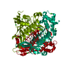
| ||||||||||||
| Unit cell |
| ||||||||||||
| Components on special symmetry positions |
|
-Components
| #1: Protein | Mass: 29966.992 Da / Num. of mol.: 1 Source method: isolated from a genetically manipulated source Source: (gene. exp.) Bordetella pertussis Tohama I (bacteria)Gene: dapD, BP1764 / Plasmid: BopeA.00002.a.B2 / Production host: References: UniProt: P0A4U8, 2,3,4,5-tetrahydropyridine-2,6-dicarboxylate N-succinyltransferase | ||||||||||
|---|---|---|---|---|---|---|---|---|---|---|---|
| #2: Chemical |   Mass: 40.078 Da / Num. of mol.: 3 / Source method: obtained synthetically / Formula: Ca Mass: 40.078 Da / Num. of mol.: 3 / Source method: obtained synthetically / Formula: Ca#3: Chemical |   Mass: 35.453 Da / Num. of mol.: 3 / Source method: obtained synthetically / Formula: Cl Mass: 35.453 Da / Num. of mol.: 3 / Source method: obtained synthetically / Formula: Cl#4: Chemical | ChemComp-GOL / |   Mass: 92.094 Da / Num. of mol.: 1 / Source method: obtained synthetically / Formula: C3H8O3 Mass: 92.094 Da / Num. of mol.: 1 / Source method: obtained synthetically / Formula: C3H8O3#5: Water | ChemComp-HOH / |  Mass: 18.015 Da / Num. of mol.: 232 / Source method: isolated from a natural source / Formula: H2O Mass: 18.015 Da / Num. of mol.: 232 / Source method: isolated from a natural source / Formula: H2OHas ligand of interest | N | Has protein modification | N | |
-Experimental details
-Experiment
| Experiment | Method: X-RAY DIFFRACTION / Number of used crystals: 1 |
|---|
- Sample preparation
| Crystal | Density Matthews: 3.6 Å3/Da / Density % sol: 65.82 % |
|---|---|
| Crystal grow | Temperature: 291 K / Method: vapor diffusion, sitting drop / pH: 4 Details: Berkeley A1: 2200 mM Sodium chloride, 100 mM Tris HCl/ Sodium hydroxide pH 8.0, 50 mM Calcium chloride. BopeA.00002.a.B2.PW39337 at 16.9 mg/mL. plate 19707 A1 drop 1, Puck: PSL1213, Cryo: ...Details: Berkeley A1: 2200 mM Sodium chloride, 100 mM Tris HCl/ Sodium hydroxide pH 8.0, 50 mM Calcium chloride. BopeA.00002.a.B2.PW39337 at 16.9 mg/mL. plate 19707 A1 drop 1, Puck: PSL1213, Cryo: 80% crystallant + 20% glycerol. |
-Data collection
| Diffraction | Mean temperature: 100 K / Serial crystal experiment: N |
|---|---|
| Diffraction source | Source: SYNCHROTRON / Site: NSLS-II / Beamline: 19-ID / Wavelength: 0.9786 Å |
| Detector | Type: DECTRIS EIGER2 XE 9M / Detector: PIXEL / Date: Mar 15, 2025 |
| Radiation | Monochromator: Double Crystal Si 111 / Protocol: SINGLE WAVELENGTH / Monochromatic (M) / Laue (L): M / Scattering type: x-ray |
| Radiation wavelength | Wavelength: 0.9786 Å / Relative weight: 1 |
| Reflection | Resolution: 1.59→48.85 Å / Num. obs: 57075 / % possible obs: 100 % / Redundancy: 20.6 % / CC1/2: 0.999 / Rmerge(I) obs: 0.196 / Rpim(I) all: 0.044 / Rrim(I) all: 0.201 / Χ2: 0.99 / Net I/σ(I): 13.3 / Num. measured all: 1173267 |
| Reflection shell | Resolution: 1.59→1.62 Å / % possible obs: 100 % / Redundancy: 21.4 % / Rmerge(I) obs: 2.87 / Num. measured all: 60173 / Num. unique obs: 2815 / CC1/2: 0.308 / Rpim(I) all: 0.63 / Rrim(I) all: 2.939 / Χ2: 0.99 / Net I/σ(I) obs: 1.6 |
- Processing
| Software |
| ||||||||||||||||||||||||||||||||||||||||||||||||||||||||||||||||||||||||||||||||||||||||||||||||||||||||||||||||||||||||||||||||||||||||||||||||||||||||||||||||||||||||||||||||||||||||||||||||||||||||||||||||||||||||||||||||||||||||||||||||||||||||||
|---|---|---|---|---|---|---|---|---|---|---|---|---|---|---|---|---|---|---|---|---|---|---|---|---|---|---|---|---|---|---|---|---|---|---|---|---|---|---|---|---|---|---|---|---|---|---|---|---|---|---|---|---|---|---|---|---|---|---|---|---|---|---|---|---|---|---|---|---|---|---|---|---|---|---|---|---|---|---|---|---|---|---|---|---|---|---|---|---|---|---|---|---|---|---|---|---|---|---|---|---|---|---|---|---|---|---|---|---|---|---|---|---|---|---|---|---|---|---|---|---|---|---|---|---|---|---|---|---|---|---|---|---|---|---|---|---|---|---|---|---|---|---|---|---|---|---|---|---|---|---|---|---|---|---|---|---|---|---|---|---|---|---|---|---|---|---|---|---|---|---|---|---|---|---|---|---|---|---|---|---|---|---|---|---|---|---|---|---|---|---|---|---|---|---|---|---|---|---|---|---|---|---|---|---|---|---|---|---|---|---|---|---|---|---|---|---|---|---|---|---|---|---|---|---|---|---|---|---|---|---|---|---|---|---|---|---|---|---|---|---|---|---|---|---|---|---|---|---|---|---|---|
| Refinement | Method to determine structure: MOLECULAR REPLACEMENT / Resolution: 1.59→48.85 Å / SU ML: 0.19 / Cross valid method: FREE R-VALUE / σ(F): 1.34 / Phase error: 18.57 / Stereochemistry target values: ML
| ||||||||||||||||||||||||||||||||||||||||||||||||||||||||||||||||||||||||||||||||||||||||||||||||||||||||||||||||||||||||||||||||||||||||||||||||||||||||||||||||||||||||||||||||||||||||||||||||||||||||||||||||||||||||||||||||||||||||||||||||||||||||||
| Solvent computation | Shrinkage radii: 0.9 Å / VDW probe radii: 1.1 Å / Solvent model: FLAT BULK SOLVENT MODEL | ||||||||||||||||||||||||||||||||||||||||||||||||||||||||||||||||||||||||||||||||||||||||||||||||||||||||||||||||||||||||||||||||||||||||||||||||||||||||||||||||||||||||||||||||||||||||||||||||||||||||||||||||||||||||||||||||||||||||||||||||||||||||||
| Refinement step | Cycle: LAST / Resolution: 1.59→48.85 Å
| ||||||||||||||||||||||||||||||||||||||||||||||||||||||||||||||||||||||||||||||||||||||||||||||||||||||||||||||||||||||||||||||||||||||||||||||||||||||||||||||||||||||||||||||||||||||||||||||||||||||||||||||||||||||||||||||||||||||||||||||||||||||||||
| Refine LS restraints |
| ||||||||||||||||||||||||||||||||||||||||||||||||||||||||||||||||||||||||||||||||||||||||||||||||||||||||||||||||||||||||||||||||||||||||||||||||||||||||||||||||||||||||||||||||||||||||||||||||||||||||||||||||||||||||||||||||||||||||||||||||||||||||||
| LS refinement shell |
| ||||||||||||||||||||||||||||||||||||||||||||||||||||||||||||||||||||||||||||||||||||||||||||||||||||||||||||||||||||||||||||||||||||||||||||||||||||||||||||||||||||||||||||||||||||||||||||||||||||||||||||||||||||||||||||||||||||||||||||||||||||||||||
| Refinement TLS params. | Method: refined / Refine-ID: X-RAY DIFFRACTION
| ||||||||||||||||||||||||||||||||||||||||||||||||||||||||||||||||||||||||||||||||||||||||||||||||||||||||||||||||||||||||||||||||||||||||||||||||||||||||||||||||||||||||||||||||||||||||||||||||||||||||||||||||||||||||||||||||||||||||||||||||||||||||||
| Refinement TLS group |
|
