 Movie
Movie Controller

[English] 日本語
 Yorodumi
- PDB-9nx4: Crystal Structure of a P. Aeruginosa Gyrase Chimera In Complex wi... -
+ Open data

- Basic information
| Entry | Database: PDB / ID: 9nx4 | ||||||||||||
|---|---|---|---|---|---|---|---|---|---|---|---|---|---|
| Title | Crystal Structure of a P. Aeruginosa Gyrase Chimera In Complex with 20mer DNA and Ciprofloxacin | ||||||||||||
 Components Components |
| ||||||||||||
 Keywords Keywords | DNA BINDING PROTEIN / Gyrase / Pseudomonas / DNA binding / Ciprofloxacin | ||||||||||||
| Function / homology |  Function and homology information Function and homology informationDNA topoisomerase type II (double strand cut, ATP-hydrolyzing) complex / DNA negative supercoiling activity / DNA topoisomerase type II (double strand cut, ATP-hydrolyzing) activity / DNA topoisomerase (ATP-hydrolysing) / DNA topological change / DNA-templated DNA replication / chromosome / DNA binding / ATP binding / metal ion binding / cytoplasm Similarity search - Function | ||||||||||||
| Biological species |   Pseudomonas aeruginosa (bacteria) Pseudomonas aeruginosa (bacteria) | ||||||||||||
| Method |  X-RAY DIFFRACTION / SYNCHROTRON / MOLECULAR REPLACEMENT / Resolution: 2.85 Å X-RAY DIFFRACTION / SYNCHROTRON / MOLECULAR REPLACEMENT / Resolution: 2.85 Å | ||||||||||||
 Authors Authors | Pedersen, L.C. / Doetsch, P.W. / Garcia-Villada, L. / Degtyareva, N. / Perera, U.L. | ||||||||||||
| Funding support |  United States, 3items United States, 3items
| ||||||||||||
 Citation Citation | Journal: Biomolecules / Year: 2026 Title: Structural and Computational Analysis of Pseudomonas aeruginosa DNA Gyrase Reveals Molecular Characteristics That May Contribute to Ciprofloxacin Resistance. Authors: Perera, L. / Garcia-Villada, L. / Kaminski, A.M. / Degtyareva, N. / Pedersen, L.C. / Doetsch, P.W. #1: Journal: Acta Crystallogr.,Sect.D / Year: 2012 Title: Towards automated crystallographic structure refinement with phenix.refine. Authors: Afonine, P.V. / Grosse-Kunstleve, R.W. / Echols, N. / Headd, J.J. / Moriarty, N.W. / Mustyakimov, M. / Terwilliger, T.C. / Urzhumtsev, A. / Zwart, P.H. / Adams, P.D. #2: Journal: Acta Crystallogr D Struct Biol / Year: 2019 Title: Macromolecular structure determination using X-rays, neutrons and electrons: recent developments in Phenix. Authors: Dorothee Liebschner / Pavel V Afonine / Matthew L Baker / Gábor Bunkóczi / Vincent B Chen / Tristan I Croll / Bradley Hintze / Li Wei Hung / Swati Jain / Airlie J McCoy / Nigel W Moriarty ...Authors: Dorothee Liebschner / Pavel V Afonine / Matthew L Baker / Gábor Bunkóczi / Vincent B Chen / Tristan I Croll / Bradley Hintze / Li Wei Hung / Swati Jain / Airlie J McCoy / Nigel W Moriarty / Robert D Oeffner / Billy K Poon / Michael G Prisant / Randy J Read / Jane S Richardson / David C Richardson / Massimo D Sammito / Oleg V Sobolev / Duncan H Stockwell / Thomas C Terwilliger / Alexandre G Urzhumtsev / Lizbeth L Videau / Christopher J Williams / Paul D Adams /   Abstract: Diffraction (X-ray, neutron and electron) and electron cryo-microscopy are powerful methods to determine three-dimensional macromolecular structures, which are required to understand biological ...Diffraction (X-ray, neutron and electron) and electron cryo-microscopy are powerful methods to determine three-dimensional macromolecular structures, which are required to understand biological processes and to develop new therapeutics against diseases. The overall structure-solution workflow is similar for these techniques, but nuances exist because the properties of the reduced experimental data are different. Software tools for structure determination should therefore be tailored for each method. Phenix is a comprehensive software package for macromolecular structure determination that handles data from any of these techniques. Tasks performed with Phenix include data-quality assessment, map improvement, model building, the validation/rebuilding/refinement cycle and deposition. Each tool caters to the type of experimental data. The design of Phenix emphasizes the automation of procedures, where possible, to minimize repetitive and time-consuming manual tasks, while default parameters are chosen to encourage best practice. A graphical user interface provides access to many command-line features of Phenix and streamlines the transition between programs, project tracking and re-running of previous tasks. | ||||||||||||
| History |
|
- Structure visualization
| Structure viewer | Molecule: MolmilJmol/JSmol |
|---|
- Downloads & links
-Download
| PDBx/mmCIF format | 9nx4.cif.gz | 509.5 KB | Display | PDBx/mmCIF format |
|---|---|---|---|---|
| PDB format | pdb9nx4.ent.gz | 372.7 KB | Display | PDB format |
| PDBx/mmJSON format | 9nx4.json.gz | Tree view | PDBx/mmJSON format | |
| Others |  Other downloads Other downloads |
-Validation report
| Arichive directory | https://data.pdbj.org/pub/pdb/validation_reports/nx/9nx4ftp://data.pdbj.org/pub/pdb/validation_reports/nx/9nx4 | HTTPS FTP |
|---|
-Related structure data
| Similar structure data | |
|---|---|
| Other databases |
|
-Links
 PDBj
PDBj


- Assembly
| Deposited unit | 
| ||||||||||||
|---|---|---|---|---|---|---|---|---|---|---|---|---|---|
| 1 | 
| ||||||||||||
| Unit cell |
|
-Components
-Protein / DNA chain , 2 types, 2 molecules AC
| #1: Protein | Mass: 105399.266 Da / Num. of mol.: 1 Source method: isolated from a genetically manipulated source Details: GSAAA(tag)-GyrB(residues 397-806)-GGS(linker)-GyrA(residues 2-525),GSAAA(tag)-GyrB(residues 397-806)-GSS(linker)-GyrA(residues 2-525) Source: (gene. exp.) Pseudomonas aeruginosa (bacteria) / Gene: gyrB, gyrA, CSB93_3133 / Production host: References: UniProt: Q9S1C7, UniProt: A0A2R3INH2, DNA topoisomerase (ATP-hydrolysing) |
|---|---|
| #2: DNA chain | Mass: 6135.955 Da / Num. of mol.: 1 / Source method: obtained synthetically / Source: (synth.) |
-Non-polymers , 6 types, 27 molecules 


| #3: Chemical | ChemComp-CIT /  Mass: 192.124 Da / Num. of mol.: 1 / Source method: obtained synthetically / Formula: C6H8O7 Mass: 192.124 Da / Num. of mol.: 1 / Source method: obtained synthetically / Formula: C6H8O7 | ||||||||
|---|---|---|---|---|---|---|---|---|---|
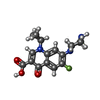
| #4: Chemical |  Mass: 331.342 Da / Num. of mol.: 2 / Source method: obtained synthetically / Formula: C17H18FN3O3 / Feature type: SUBJECT OF INVESTIGATION / Comment: antibiotic*YM Mass: 331.342 Da / Num. of mol.: 2 / Source method: obtained synthetically / Formula: C17H18FN3O3 / Feature type: SUBJECT OF INVESTIGATION / Comment: antibiotic*YM#5: Chemical | ChemComp-SO4 /  Mass: 96.063 Da / Num. of mol.: 4 / Source method: obtained synthetically / Formula: SO4 Mass: 96.063 Da / Num. of mol.: 4 / Source method: obtained synthetically / Formula: SO4#6: Chemical | ChemComp-MN / |  Mass: 54.938 Da / Num. of mol.: 1 / Source method: obtained synthetically / Formula: Mn Mass: 54.938 Da / Num. of mol.: 1 / Source method: obtained synthetically / Formula: Mn#7: Chemical |  Mass: 35.453 Da / Num. of mol.: 3 / Source method: obtained synthetically / Formula: Cl Mass: 35.453 Da / Num. of mol.: 3 / Source method: obtained synthetically / Formula: Cl#8: Water | ChemComp-HOH / | Mass: 18.015 Da / Num. of mol.: 16 / Source method: isolated from a natural source / Formula: H2O |
-Details
| Has ligand of interest | Y |
|---|---|
| Has protein modification | N |
-Experimental details
-Experiment
| Experiment | Method: X-RAY DIFFRACTION / Number of used crystals: 1 |
|---|
- Sample preparation
| Crystal | Density Matthews: 3.39 Å3/Da / Density % sol: 63.68 % |
|---|---|
| Crystal grow | Temperature: 294 K / Method: vapor diffusion, hanging drop / pH: 6 Details: 0.1M sodium citrate pH 6.0 14% PEG 4000 200mM Ammonium Sulfate 1mM MnCl2 1mM ciprofloxacin Temp details: room termperature |
-Data collection
| Diffraction | Mean temperature: 100 K / Serial crystal experiment: N |
|---|---|
| Diffraction source | Source: SYNCHROTRON / Site: APS / Beamline: 22-ID / Wavelength: 1 Å |
| Detector | Type: DECTRIS EIGER X 16M / Detector: PIXEL / Date: Oct 8, 2021 |
| Radiation | Protocol: SINGLE WAVELENGTH / Monochromatic (M) / Laue (L): M / Scattering type: x-ray |
| Radiation wavelength | Wavelength: 1 Å / Relative weight: 1 |
| Reflection | Resolution: 2.85→50 Å / Num. obs: 36343 / % possible obs: 99.2 % / Redundancy: 15.5 % / Biso Wilson estimate: 51.72 Å2 / CC1/2: 0.992 / Rmerge(I) obs: 0.168 / Rpim(I) all: 0.043 / Rrim(I) all: 0.174 / Net I/σ(I): 3.7 |
| Reflection shell | Resolution: 2.85→2.9 Å / Rmerge(I) obs: 1.252 / Mean I/σ(I) obs: 1.29 / Num. unique obs: 1785 / CC1/2: 0.815 / Rpim(I) all: 0.327 / Rrim(I) all: 1.296 |
- Processing
| Software |
| |||||||||||||||||||||||||||||||||||||||||||||||||||||||||||||||||||||||||||||||||||||||||||||||||||||||||||||||||||||||||||||
|---|---|---|---|---|---|---|---|---|---|---|---|---|---|---|---|---|---|---|---|---|---|---|---|---|---|---|---|---|---|---|---|---|---|---|---|---|---|---|---|---|---|---|---|---|---|---|---|---|---|---|---|---|---|---|---|---|---|---|---|---|---|---|---|---|---|---|---|---|---|---|---|---|---|---|---|---|---|---|---|---|---|---|---|---|---|---|---|---|---|---|---|---|---|---|---|---|---|---|---|---|---|---|---|---|---|---|---|---|---|---|---|---|---|---|---|---|---|---|---|---|---|---|---|---|---|---|
| Refinement | Method to determine structure: MOLECULAR REPLACEMENT / Resolution: 2.85→43.67 Å / SU ML: 0.3688 / Cross valid method: FREE R-VALUE / σ(F): 1.35 / Phase error: 25.2092 Stereochemistry target values: GeoStd + Monomer Library + CDL v1.2
| |||||||||||||||||||||||||||||||||||||||||||||||||||||||||||||||||||||||||||||||||||||||||||||||||||||||||||||||||||||||||||||
| Solvent computation | Shrinkage radii: 0.9 Å / VDW probe radii: 1.11 Å / Solvent model: FLAT BULK SOLVENT MODEL | |||||||||||||||||||||||||||||||||||||||||||||||||||||||||||||||||||||||||||||||||||||||||||||||||||||||||||||||||||||||||||||
| Displacement parameters | Biso mean: 58.3 Å2 | |||||||||||||||||||||||||||||||||||||||||||||||||||||||||||||||||||||||||||||||||||||||||||||||||||||||||||||||||||||||||||||
| Refinement step | Cycle: LAST / Resolution: 2.85→43.67 Å
| |||||||||||||||||||||||||||||||||||||||||||||||||||||||||||||||||||||||||||||||||||||||||||||||||||||||||||||||||||||||||||||
| Refine LS restraints |
| |||||||||||||||||||||||||||||||||||||||||||||||||||||||||||||||||||||||||||||||||||||||||||||||||||||||||||||||||||||||||||||
| LS refinement shell |
| |||||||||||||||||||||||||||||||||||||||||||||||||||||||||||||||||||||||||||||||||||||||||||||||||||||||||||||||||||||||||||||
| Refinement TLS params. | Method: refined / Refine-ID: X-RAY DIFFRACTION
| |||||||||||||||||||||||||||||||||||||||||||||||||||||||||||||||||||||||||||||||||||||||||||||||||||||||||||||||||||||||||||||
| Refinement TLS group | Refine-ID: X-RAY DIFFRACTION
|
