 Movie
Movie Controller

[English] 日本語
 Yorodumi
- PDB-9nr5: Crystal structure of H5 hemagglutinin Q226L mutant from the influ... -
+ Open data

- Basic information
| Entry | Database: PDB / ID: 9nr5 | ||||||
|---|---|---|---|---|---|---|---|
| Title | Crystal structure of H5 hemagglutinin Q226L mutant from the influenza virus A/black swan/Akita/1/2016 with LSTc | ||||||
 Components Components | (Hemagglutinin ...) x 2 | ||||||
 Keywords Keywords | VIRAL PROTEIN / H1N1 / Antibody / Hemagglutinin | ||||||
| Function / homology |  Function and homology information Function and homology informationviral budding from plasma membrane / clathrin-dependent endocytosis of virus by host cell / host cell surface receptor binding / fusion of virus membrane with host plasma membrane / fusion of virus membrane with host endosome membrane / viral envelope / virion attachment to host cell / host cell plasma membrane / virion membrane / membrane Similarity search - Function | ||||||
| Biological species |   Influenza A virus Influenza A virus | ||||||
| Method |  X-RAY DIFFRACTION / SYNCHROTRON / MOLECULAR REPLACEMENT / Resolution: 2.52 Å X-RAY DIFFRACTION / SYNCHROTRON / MOLECULAR REPLACEMENT / Resolution: 2.52 Å | ||||||
 Authors Authors | Lin, T.H. / Zhu, Y. / Wilson, I.A. | ||||||
| Funding support |  United States, 1items United States, 1items
| ||||||
 Citation Citation | Journal: Proc.Natl.Acad.Sci.USA / Year: 2025 Title: The Q226L mutation can convert a highly pathogenic H5 2.3.4.4e virus to bind human-type receptors. Authors: Rios Carrasco, M. / Lin, T.H. / Zhu, X. / Gabarroca Garcia, A. / Uslu, E. / Liang, R. / Spruit, C.M. / Richard, M. / Boons, G.J. / Wilson, I.A. / de Vries, R.P. | ||||||
| History |
|
- Structure visualization
| Structure viewer | Molecule: MolmilJmol/JSmol |
|---|
- Downloads & links
-Download
| PDBx/mmCIF format | 9nr5.cif.gz | 311 KB | Display | PDBx/mmCIF format |
|---|---|---|---|---|
| PDB format | pdb9nr5.ent.gz | 251.5 KB | Display | PDB format |
| PDBx/mmJSON format | 9nr5.json.gz | Tree view | PDBx/mmJSON format | |
| Others |  Other downloads Other downloads |
-Validation report
| Summary document | 9nr5_validation.pdf.gz | 3.9 MB | Display | wwPDB validaton report |
|---|---|---|---|---|
| Full document | 9nr5_full_validation.pdf.gz | 4 MB | Display | |
| Data in XML | 9nr5_validation.xml.gz | 31.7 KB | Display | |
| Data in CIF | 9nr5_validation.cif.gz | 47.1 KB | Display | |
| Arichive directory | https://data.pdbj.org/pub/pdb/validation_reports/nr/9nr5ftp://data.pdbj.org/pub/pdb/validation_reports/nr/9nr5 | HTTPS FTP |
-Related structure data


-Links
 PDBj
PDBj


- Assembly
| Deposited unit | 
| ||||||||
|---|---|---|---|---|---|---|---|---|---|
| 1 |
| ||||||||
| Unit cell |
|
-Components
-Hemagglutinin ... , 2 types, 6 molecules ACEBDF
| #1: Protein | Mass: 36509.453 Da / Num. of mol.: 3 / Mutation: Q226L Source method: isolated from a genetically manipulated source Source: (gene. exp.) Influenza A virus / Gene: HAProduction host:  Spodoptera aff. frugiperda 1 BOLD-2017 (butterflies/moths) Spodoptera aff. frugiperda 1 BOLD-2017 (butterflies/moths)References: UniProt: A0A1L7N0F8 #2: Protein | Mass: 20442.520 Da / Num. of mol.: 3 Source method: isolated from a genetically manipulated source Source: (gene. exp.) Influenza A virus / Gene: HAProduction host: Spodoptera aff. frugiperda 1 BOLD-2017 (butterflies/moths)References: UniProt: A0A1L7N0F8 |
|---|
-Sugars , 4 types, 13 molecules 
| #3: Polysaccharide | Source method: isolated from a genetically manipulated source #4: Polysaccharide | beta-D-mannopyranose-(1-4)-2-acetamido-2-deoxy-beta-D-glucopyranose-(1-4)-2-acetamido-2-deoxy-beta- ...beta-D-mannopyranose-(1-4)-2-acetamido-2-deoxy-beta-D-glucopyranose-(1-4)-2-acetamido-2-deoxy-beta-D-glucopyranose | Source method: isolated from a genetically manipulated source #5: Polysaccharide | 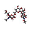  Source method: isolated from a genetically manipulated source Details: oligosaccharide / References: 6'-sialyl-N-acetyllactosamine #6: Sugar | ChemComp-NAG /  Type: D-saccharide, beta linking / Mass: 221.208 Da / Num. of mol.: 6 / Source method: obtained synthetically / Formula: C8H15NO6 / Feature type: SUBJECT OF INVESTIGATION Type: D-saccharide, beta linking / Mass: 221.208 Da / Num. of mol.: 6 / Source method: obtained synthetically / Formula: C8H15NO6 / Feature type: SUBJECT OF INVESTIGATION |
|---|
-Non-polymers , 1 types, 56 molecules 
| #7: Water | ChemComp-HOH / Mass: 18.015 Da / Num. of mol.: 56 / Source method: isolated from a natural source / Formula: H2O |
|---|
-Details
| Has ligand of interest | Y |
|---|---|
| Has protein modification | Y |
-Experimental details
-Experiment
| Experiment | Method: X-RAY DIFFRACTION / Number of used crystals: 1 |
|---|
- Sample preparation
| Crystal | Density Matthews: 2.93 Å3/Da / Density % sol: 57.99 % |
|---|---|
| Crystal grow | Temperature: 293 K / Method: vapor diffusion, hanging drop / Details: 0.2M ammonium format, 20% PEG3350 |
-Data collection
| Diffraction | Mean temperature: 100 K / Serial crystal experiment: N |
|---|---|
| Diffraction source | Source: SYNCHROTRON / Site: SSRL / Beamline: BL12-1 / Wavelength: 0.979 Å |
| Detector | Type: DECTRIS EIGER X 16M / Detector: PIXEL / Date: Jan 31, 2024 |
| Radiation | Protocol: SINGLE WAVELENGTH / Monochromatic (M) / Laue (L): M / Scattering type: x-ray |
| Radiation wavelength | Wavelength: 0.979 Å / Relative weight: 1 |
| Reflection | Resolution: 2.52→39.68 Å / Num. obs: 65184 / % possible obs: 98 % / Redundancy: 6.8 % / CC1/2: 0.99 / Rpim(I) all: 0.049 / Rrim(I) all: 0.127 / Net I/σ(I): 23.8 |
| Reflection shell | Resolution: 2.52→2.56 Å / Num. unique obs: 6297 / CC1/2: 0.78 / Rpim(I) all: 0.36 / Rrim(I) all: 0.95 |
- Processing
| Software |
| ||||||||||||||||||||||||||||||||||||||||||||||||||||||||||||||||||||||||||||||||||||||||||||||||||||||||||||||||||||||||||||||||||||||||||||||||||||||||||||||||||||||||
|---|---|---|---|---|---|---|---|---|---|---|---|---|---|---|---|---|---|---|---|---|---|---|---|---|---|---|---|---|---|---|---|---|---|---|---|---|---|---|---|---|---|---|---|---|---|---|---|---|---|---|---|---|---|---|---|---|---|---|---|---|---|---|---|---|---|---|---|---|---|---|---|---|---|---|---|---|---|---|---|---|---|---|---|---|---|---|---|---|---|---|---|---|---|---|---|---|---|---|---|---|---|---|---|---|---|---|---|---|---|---|---|---|---|---|---|---|---|---|---|---|---|---|---|---|---|---|---|---|---|---|---|---|---|---|---|---|---|---|---|---|---|---|---|---|---|---|---|---|---|---|---|---|---|---|---|---|---|---|---|---|---|---|---|---|---|---|---|---|---|
| Refinement | Method to determine structure: MOLECULAR REPLACEMENT / Resolution: 2.52→39.68 Å / SU ML: 0.38 / Cross valid method: FREE R-VALUE / σ(F): 1.37 / Phase error: 28.72 / Stereochemistry target values: ML
| ||||||||||||||||||||||||||||||||||||||||||||||||||||||||||||||||||||||||||||||||||||||||||||||||||||||||||||||||||||||||||||||||||||||||||||||||||||||||||||||||||||||||
| Solvent computation | Shrinkage radii: 0.9 Å / VDW probe radii: 1.1 Å / Solvent model: FLAT BULK SOLVENT MODEL | ||||||||||||||||||||||||||||||||||||||||||||||||||||||||||||||||||||||||||||||||||||||||||||||||||||||||||||||||||||||||||||||||||||||||||||||||||||||||||||||||||||||||
| Refinement step | Cycle: LAST / Resolution: 2.52→39.68 Å
| ||||||||||||||||||||||||||||||||||||||||||||||||||||||||||||||||||||||||||||||||||||||||||||||||||||||||||||||||||||||||||||||||||||||||||||||||||||||||||||||||||||||||
| Refine LS restraints |
| ||||||||||||||||||||||||||||||||||||||||||||||||||||||||||||||||||||||||||||||||||||||||||||||||||||||||||||||||||||||||||||||||||||||||||||||||||||||||||||||||||||||||
| LS refinement shell |
|
