 Movie
Movie Controller


+ Open data

- Basic information
| Entry | Database: PDB / ID: 9njs | |||||||||
|---|---|---|---|---|---|---|---|---|---|---|
| Title | human polycystin-2 with clinical variant D511V | |||||||||
 Components Components | Polycystin-2 | |||||||||
 Keywords Keywords | METAL TRANSPORT / ion channels / polycystic kidney disease / PKD / PKD2 / D511V / TRP / ADPKD | |||||||||
| Function / homology |  Function and homology information Function and homology informationdetection of nodal flow / metanephric smooth muscle tissue development / metanephric cortex development / metanephric cortical collecting duct development / metanephric distal tubule development / polycystin complex / mesonephric tubule development / mesonephric duct development / metanephric part of ureteric bud development / renal tubule morphogenesis ...detection of nodal flow / metanephric smooth muscle tissue development / metanephric cortex development / metanephric cortical collecting duct development / metanephric distal tubule development / polycystin complex / mesonephric tubule development / mesonephric duct development / metanephric part of ureteric bud development / renal tubule morphogenesis / determination of liver left/right asymmetry / metanephric ascending thin limb development / metanephric mesenchyme development / metanephric S-shaped body morphogenesis / basal cortex / placenta blood vessel development / renal artery morphogenesis / HLH domain binding / VxPx cargo-targeting to cilium / cilium organization / migrasome / neural tube development / cellular response to fluid shear stress / regulation of calcium ion import / calcium-induced calcium release activity / detection of mechanical stimulus / determination of left/right symmetry / voltage-gated monoatomic ion channel activity / embryonic placenta development / cellular response to hydrostatic pressure / aorta development / cation channel complex / branching involved in ureteric bud morphogenesis / non-motile cilium / outward rectifier potassium channel activity / motile cilium / actinin binding / cellular response to osmotic stress / negative regulation of G1/S transition of mitotic cell cycle / voltage-gated sodium channel activity / heart looping / spinal cord development / ciliary membrane / voltage-gated monoatomic cation channel activity / positive regulation of phospholipase C-activating G protein-coupled receptor signaling pathway / protein heterotetramerization / cytoplasmic side of endoplasmic reticulum membrane / potassium channel activity / voltage-gated potassium channel activity / centrosome duplication / transcription regulator inhibitor activity / cell surface receptor signaling pathway via JAK-STAT / voltage-gated calcium channel activity / release of sequestered calcium ion into cytosol / monoatomic cation channel activity / cytoskeletal protein binding / cellular response to calcium ion / cellular response to cAMP / liver development / potassium ion transmembrane transport / basal plasma membrane / cytoplasmic vesicle membrane / sodium ion transmembrane transport / lumenal side of endoplasmic reticulum membrane / cellular response to reactive oxygen species / protein tetramerization / Wnt signaling pathway / phosphoprotein binding / mitotic spindle / positive regulation of nitric oxide biosynthetic process / calcium ion transmembrane transport / heart development / calcium ion transport / transmembrane transport / cell-cell junction / cilium / regulation of cell population proliferation / lamellipodium / ATPase binding / ciliary basal body / vesicle / protein homotetramerization / basolateral plasma membrane / regulation of cell cycle / transmembrane transporter binding / cell surface receptor signaling pathway / negative regulation of cell population proliferation / signaling receptor binding / positive regulation of gene expression / calcium ion binding / endoplasmic reticulum membrane / Golgi apparatus / endoplasmic reticulum / positive regulation of transcription by RNA polymerase II / protein homodimerization activity / extracellular exosome / membrane / identical protein binding / plasma membrane / cytoplasm Similarity search - Function | |||||||||
| Biological species |  Homo sapiens (human) Homo sapiens (human) | |||||||||
| Method | ELECTRON MICROSCOPY / single particle reconstruction / cryo EM / Resolution: 3.1 Å | |||||||||
 Authors Authors | Wang, Q. / Cao, E. | |||||||||
| Funding support |  United States, 1items United States, 1items
| |||||||||
 Citation Citation | Journal: To Be Published Title: human polycystin-2 with clinical variant D511V Authors: Wang, Q. / Cao, E. | |||||||||
| History |
|
- Structure visualization
| Structure viewer | Molecule: MolmilJmol/JSmol |
|---|
- Downloads & links
-Download
| PDBx/mmCIF format | 9njs.cif.gz | 396.5 KB | Display | PDBx/mmCIF format |
|---|---|---|---|---|
| PDB format | pdb9njs.ent.gz | 317.1 KB | Display | PDB format |
| PDBx/mmJSON format | 9njs.json.gz | Tree view | PDBx/mmJSON format | |
| Others |  Other downloads Other downloads |
-Validation report
| Arichive directory | https://data.pdbj.org/pub/pdb/validation_reports/nj/9njsftp://data.pdbj.org/pub/pdb/validation_reports/nj/9njs | HTTPS FTP |
|---|
-Related structure data
| Related structure data |  49489MC M: map data used to model this data C: citing same article ( |
|---|---|
| Similar structure data |
-Links
 PDBj
PDBj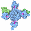
- Assembly
| Deposited unit | 
|
|---|---|
| 1 |
|
-Components
| #1: Protein | Mass: 85433.898 Da / Num. of mol.: 4 / Mutation: D511V Source method: isolated from a genetically manipulated source Details: human polycystin-2 53-792 with clinical variant D511V Source: (gene. exp.) Homo sapiens (human) / Gene: PKD2, TRPP1, TRPP2 / Production host: Homo sapiens (human) / References: UniProt: Q13563#2: Sugar | ChemComp-NAG /   Type: D-saccharide, beta linking / Mass: 221.208 Da / Num. of mol.: 12 Type: D-saccharide, beta linking / Mass: 221.208 Da / Num. of mol.: 12Source method: isolated from a genetically manipulated source Formula: C8H15NO6 Has ligand of interest | N | Has protein modification | Y | |
|---|
-Experimental details
-Experiment
| Experiment | Method: ELECTRON MICROSCOPY |
|---|---|
| EM experiment | Aggregation state: PARTICLE / 3D reconstruction method: single particle reconstruction |
- Sample preparation
| Component | Name: human polycystin-2 53-792 with clinical variant D511V / Type: COMPLEX Details: Over expressed in HEK293S cells and purified in detergent Entity ID: #1 / Source: RECOMBINANT |
|---|---|
| Molecular weight | Experimental value: NO |
| Source (natural) | Organism: Homo sapiens (human) |
| Source (recombinant) | Organism: Homo sapiens (human) / Cell: HEK293 |
| Buffer solution | pH: 7.4 |
| Specimen | Embedding applied: NO / Shadowing applied: NO / Staining applied: NO / Vitrification applied: YES |
| Vitrification | Instrument: FEI VITROBOT MARK III / Cryogen name: ETHANE |
- Electron microscopy imaging
| Experimental equipment |  Model: Titan Krios / Image courtesy: FEI Company |
|---|---|
| Microscopy | Model: TFS KRIOS |
| Electron gun | Electron source:  FIELD EMISSION GUN / Accelerating voltage: 300 kV / Illumination mode: FLOOD BEAM FIELD EMISSION GUN / Accelerating voltage: 300 kV / Illumination mode: FLOOD BEAM |
| Electron lens | Mode: BRIGHT FIELD / Nominal defocus max: 4000 nm / Nominal defocus min: 1000 nm |
| Image recording | Electron dose: 59 e/Å2 / Film or detector model: GATAN K2 SUMMIT (4k x 4k) |
- Processing
| EM software | Name: PHENIX / Version: 1.20.1_4487 / Category: model refinement |
|---|---|
| CTF correction | Type: PHASE FLIPPING AND AMPLITUDE CORRECTION |
| Symmetry | Point symmetry: C4 (4 fold cyclic) |
| 3D reconstruction | Resolution: 3.1 Å / Resolution method: FSC 0.143 CUT-OFF / Num. of particles: 54727 / Symmetry type: POINT |
| Refinement | Highest resolution: 3.1 Å Stereochemistry target values: REAL-SPACE (WEIGHTED MAP SUM AT ATOM CENTERS) |
