Movie
Movie Controller

[English] 日本語
 Yorodumi
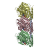
- PDB-9n9g: Cryo-EM structure of the human Hec1-Nuf2 dimer bound to the pacli... -
+ Open data

- Basic information
| Entry | Database: PDB / ID: 9n9g | |||||||||||||||||||||||||||
|---|---|---|---|---|---|---|---|---|---|---|---|---|---|---|---|---|---|---|---|---|---|---|---|---|---|---|---|---|
| Title | Cryo-EM structure of the human Hec1-Nuf2 dimer bound to the paclitaxel-stabilized microtubule | |||||||||||||||||||||||||||
 Components Components |
| |||||||||||||||||||||||||||
 Keywords Keywords | CELL CYCLE / microtubule / kinetochore / Ndc80 complex / mitosis | |||||||||||||||||||||||||||
| Function / homology |  Function and homology information Function and homology informationG2/MI transition of meiotic cell cycle / kinetochore adaptor activity / skeletal muscle satellite cell proliferation / Ndc80 complex / kinetochore organization / meiotic chromosome segregation / metaphase chromosome alignment / attachment of spindle microtubules to kinetochore / positive regulation of mitotic cell cycle spindle assembly checkpoint / spindle assembly involved in female meiosis I ...G2/MI transition of meiotic cell cycle / kinetochore adaptor activity / skeletal muscle satellite cell proliferation / Ndc80 complex / kinetochore organization / meiotic chromosome segregation / metaphase chromosome alignment / attachment of spindle microtubules to kinetochore / positive regulation of mitotic cell cycle spindle assembly checkpoint / spindle assembly involved in female meiosis I / outer kinetochore / attachment of mitotic spindle microtubules to kinetochore / Microtubule-dependent trafficking of connexons from Golgi to the plasma membrane / Resolution of Sister Chromatid Cohesion / Hedgehog 'off' state / Cilium Assembly / Intraflagellar transport / COPI-dependent Golgi-to-ER retrograde traffic / Mitotic Prometaphase / Carboxyterminal post-translational modifications of tubulin / RHOH GTPase cycle / EML4 and NUDC in mitotic spindle formation / Sealing of the nuclear envelope (NE) by ESCRT-III / Kinesins / PKR-mediated signaling / Separation of Sister Chromatids / The role of GTSE1 in G2/M progression after G2 checkpoint / Aggrephagy / RHO GTPases activate IQGAPs / RHO GTPases Activate Formins / HSP90 chaperone cycle for steroid hormone receptors (SHR) in the presence of ligand / MHC class II antigen presentation / Recruitment of NuMA to mitotic centrosomes / COPI-mediated anterograde transport / mitotic spindle assembly checkpoint signaling / establishment of mitotic spindle orientation / centrosome duplication / mitotic sister chromatid segregation / chromosome, centromeric region / Amplification of signal from unattached kinetochores via a MAD2 inhibitory signal / Mitotic Prometaphase / EML4 and NUDC in mitotic spindle formation / cyclin binding / Resolution of Sister Chromatid Cohesion / mitotic spindle organization / chromosome segregation / regulation of protein stability / RHO GTPases Activate Formins / structural constituent of cytoskeleton / kinetochore / microtubule cytoskeleton organization / neuron migration / Separation of Sister Chromatids / mitotic cell cycle / microtubule cytoskeleton / microtubule binding / microtubule / Hydrolases; Acting on acid anhydrides; Acting on GTP to facilitate cellular and subcellular movement / nuclear speck / cell division / GTPase activity / centrosome / GTP binding / protein-containing complex binding / nucleoplasm / membrane / metal ion binding / identical protein binding / nucleus / cytoplasm / cytosol Similarity search - Function | |||||||||||||||||||||||||||
| Biological species |  Homo sapiens (human) Homo sapiens (human) | |||||||||||||||||||||||||||
| Method | ELECTRON MICROSCOPY / single particle reconstruction / cryo EM / Resolution: 3 Å | |||||||||||||||||||||||||||
 Authors Authors | Funabiki, H. / Niu, Y. | |||||||||||||||||||||||||||
| Funding support |  United States, 1items United States, 1items
| |||||||||||||||||||||||||||
 Citation Citation | Journal: Sci Adv / Year: 2026 Title: Cryo-EM structure of the human Hec1-Nuf2 dimer bound to the paclitaxel-stabilized microtubule Authors: Funabiki, H. / Niu, Y. | |||||||||||||||||||||||||||
| History |
|
- Structure visualization
| Structure viewer | Molecule: MolmilJmol/JSmol |
|---|
- Downloads & links
-Download
| PDBx/mmCIF format | 9n9g.cif.gz | 564.9 KB | Display | PDBx/mmCIF format |
|---|---|---|---|---|
| PDB format | pdb9n9g.ent.gz | 432.6 KB | Display | PDB format |
| PDBx/mmJSON format | 9n9g.json.gz | Tree view | PDBx/mmJSON format | |
| Others |  Other downloads Other downloads |
-Validation report
| Arichive directory | https://data.pdbj.org/pub/pdb/validation_reports/n9/9n9gftp://data.pdbj.org/pub/pdb/validation_reports/n9/9n9g | HTTPS FTP |
|---|
-Related structure data
| Related structure data |  49168MC  9n9eC  9n9fC  9n9hC  9n9iC  9yxqC C: citing same article ( M: map data used to model this data |
|---|---|
| Similar structure data |


-Links
 PDBj
PDBj


- Assembly
| Deposited unit | 
|
|---|---|
| 1 |
|
-Components
-Kinetochore protein ... , 2 types, 5 molecules 1NnFf
| #1: Protein | Mass: 74247.945 Da / Num. of mol.: 3 Source method: isolated from a genetically manipulated source Source: (gene. exp.) Homo sapiens (human) / Gene: NDC80, HEC, HEC1, KNTC2 / Production host:  #4: Protein | Mass: 54619.875 Da / Num. of mol.: 2 Source method: isolated from a genetically manipulated source Source: (gene. exp.) Homo sapiens (human) / Gene: NUF2, CDCA1, NUF2R / Production host: |
|---|
-Protein , 2 types, 4 molecules AaBb
| #2: Protein | Mass: 50204.445 Da / Num. of mol.: 2 / Source method: isolated from a natural source / Source: (natural) #3: Protein | Mass: 49907.770 Da / Num. of mol.: 2 / Source method: isolated from a natural source / Source: (natural) |
|---|
-Non-polymers , 4 types, 10 molecules 


| #5: Chemical |  Mass: 523.180 Da / Num. of mol.: 2 / Source method: obtained synthetically / Formula: C10H16N5O14P3 / Comment: GTP, energy-carrying molecule*YM Mass: 523.180 Da / Num. of mol.: 2 / Source method: obtained synthetically / Formula: C10H16N5O14P3 / Comment: GTP, energy-carrying molecule*YM#6: Chemical | ChemComp-MG /  Mass: 24.305 Da / Num. of mol.: 4 / Source method: obtained synthetically / Formula: Mg Mass: 24.305 Da / Num. of mol.: 4 / Source method: obtained synthetically / Formula: Mg#7: Chemical |  Type: RNA linking / Mass: 443.201 Da / Num. of mol.: 2 / Source method: obtained synthetically / Formula: C10H15N5O11P2 / Comment: GDP, energy-carrying molecule*YM Type: RNA linking / Mass: 443.201 Da / Num. of mol.: 2 / Source method: obtained synthetically / Formula: C10H15N5O11P2 / Comment: GDP, energy-carrying molecule*YM#8: Chemical |  Mass: 853.906 Da / Num. of mol.: 2 / Source method: obtained synthetically / Formula: C47H51NO14 / Comment: medication*YM Mass: 853.906 Da / Num. of mol.: 2 / Source method: obtained synthetically / Formula: C47H51NO14 / Comment: medication*YM |
|---|
-Details
| Has ligand of interest | N |
|---|---|
| Has protein modification | Y |
-Experimental details
-Experiment
| Experiment | Method: ELECTRON MICROSCOPY |
|---|---|
| EM experiment | Aggregation state: FILAMENT / 3D reconstruction method: single particle reconstruction |
- Sample preparation
| Component | Name: The human Hec1-Nuf2 dimer bound to the paclitaxel-stabilized microtubule Type: COMPLEX / Entity ID: #1-#4 / Source: RECOMBINANT |
|---|---|
| Molecular weight | Value: 4.3 kDa/nm / Experimental value: NO |
| Source (natural) | Organism: Homo sapiens (human) |
| Source (recombinant) | Organism: |
| Buffer solution | pH: 6.8 |
| Specimen | Embedding applied: NO / Shadowing applied: NO / Staining applied: NO / Vitrification applied: YES |
| Specimen support | Grid material: GOLD / Grid mesh size: 400 divisions/in. / Grid type: Quantifoil |
| Vitrification | Instrument: FEI VITROBOT MARK IV / Cryogen name: ETHANE / Humidity: 100 % / Chamber temperature: 298 K |
- Electron microscopy imaging
| Experimental equipment |  Model: Titan Krios / Image courtesy: FEI Company |
|---|---|
| Microscopy | Model: TFS KRIOS |
| Electron gun | Electron source:  FIELD EMISSION GUN / Accelerating voltage: 300 kV / Illumination mode: FLOOD BEAM FIELD EMISSION GUN / Accelerating voltage: 300 kV / Illumination mode: FLOOD BEAM |
| Electron lens | Mode: BRIGHT FIELD / Nominal defocus max: 2500 nm / Nominal defocus min: 1200 nm |
| Image recording | Electron dose: 54.6 e/Å2 / Film or detector model: GATAN K3 (6k x 4k) |
- Processing
| EM software |
| |||||||||||||||||||||||||||||||||||||||||||||||||||||||||||||||||||||||||||||||||||||||||||||||||||
|---|---|---|---|---|---|---|---|---|---|---|---|---|---|---|---|---|---|---|---|---|---|---|---|---|---|---|---|---|---|---|---|---|---|---|---|---|---|---|---|---|---|---|---|---|---|---|---|---|---|---|---|---|---|---|---|---|---|---|---|---|---|---|---|---|---|---|---|---|---|---|---|---|---|---|---|---|---|---|---|---|---|---|---|---|---|---|---|---|---|---|---|---|---|---|---|---|---|---|---|---|
| CTF correction | Type: PHASE FLIPPING AND AMPLITUDE CORRECTION | |||||||||||||||||||||||||||||||||||||||||||||||||||||||||||||||||||||||||||||||||||||||||||||||||||
| 3D reconstruction | Resolution: 3 Å / Resolution method: FSC 0.143 CUT-OFF / Num. of particles: 284470 / Symmetry type: POINT | |||||||||||||||||||||||||||||||||||||||||||||||||||||||||||||||||||||||||||||||||||||||||||||||||||
| Atomic model building |
| |||||||||||||||||||||||||||||||||||||||||||||||||||||||||||||||||||||||||||||||||||||||||||||||||||
| Atomic model building |
|