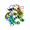
Movie
Movie Controller

[English] 日本語
 Yorodumi
- PDB-9n0h: Structure of proteinase K from energy-filtered MicroED data using... -
+ Open data

- Basic information
| Entry | Database: PDB / ID: 9n0h | |||||||||||||||
|---|---|---|---|---|---|---|---|---|---|---|---|---|---|---|---|---|
| Title | Structure of proteinase K from energy-filtered MicroED data using a 20 eV slit width | |||||||||||||||
 Components Components | Proteinase K | |||||||||||||||
 Keywords Keywords | HYDROLASE / serine protease | |||||||||||||||
| Function / homology |  Function and homology information Function and homology informationpeptidase K / serine-type endopeptidase activity / proteolysis / extracellular region Similarity search - Function | |||||||||||||||
| Biological species |  Parengyodontium album (fungus) Parengyodontium album (fungus) | |||||||||||||||
| Method | ELECTRON CRYSTALLOGRAPHY / electron crystallography / cryo EM / Resolution: 1.2 Å | |||||||||||||||
 Authors Authors | Clabbers, M.T.B. / Hattne, J. / Martynowycz, M.W. / Gonen, T. | |||||||||||||||
| Funding support |  United States, 3items United States, 3items
| |||||||||||||||
 Citation Citation | Journal: J Struct Biol X / Year: 2026 Title: Characterization of energy filtering slit widths for MicroED data collection. Authors: Max T B Clabbers / Johan Hattne / Michael W Martynowycz / Tamir Gonen / Abstract: A favorable signal-to-noise ratio is essential for obtaining high-quality diffraction data in macromolecular electron crystallography. Inelastic scattering contributes significantly to the noise, ...A favorable signal-to-noise ratio is essential for obtaining high-quality diffraction data in macromolecular electron crystallography. Inelastic scattering contributes significantly to the noise, reducing contrast between diffraction peaks and background, which complicates peak detection and compromises the accuracy of intensity integration. Energy filtering mitigates these challenges and enhances diffraction data quality by removing inelastically scattered electrons, leading to reduced background noise and sharper Bragg peaks. Previously, we reported a substantial improvement in MicroED data quality and resolution with energy filtering. Here, we systematically evaluate the impact of different energy filter slit widths for optimal MicroED data collection. Data from proteinase K lamellae were collected using 5, 10, and 20 eV energy filter slits. Our results show that the optimized energy filtering reduces background counts and improves the signal-to-noise ratio, enhancing the precision of intensity measurements and resulting in improved structural models. These findings provide insight into the optimization of energy filter slit settings that, when paired with direct electron detection, enhance data collection strategies by improving the signal-to-noise ratio, supporting higher quality data, and ultimately enabling more precise structure determination. #1: Journal: bioRxiv / Year: 2025 Title: Characterization of energy filtering slit widths for MicroED data collection. Authors: Max T B Clabbers / Johan Hattne / Michael W Martynowycz / Tamir Gonen / Abstract: A favorable signal-to-noise ratio is essential for obtaining high-quality diffraction data in macromolecular electron crystallography. Inelastic scattering contributes significantly to the noise, ...A favorable signal-to-noise ratio is essential for obtaining high-quality diffraction data in macromolecular electron crystallography. Inelastic scattering contributes significantly to the noise, reducing contrast between diffraction peaks and background, which complicates peak detection and compromises the accuracy of intensity integration. Energy filtering mitigates these challenges and enhances diffraction data quality by removing the inelastically scattered electrons, leading to reduced background noise and sharper Bragg peaks. Previously, we reported a substantial improvement in MicroED data quality and resolution with energy filtering. Here, we systematically evaluate the impact of different energy filter slit widths for optimal MicroED data collection. Data from proteinase K lamellae were collected using the 5, 10, and 20 eV energy filter slit widths. Our results show that the narrowest slit widths result in a stronger diffraction signal with lower background noise, improving the precision of the intensity measurements which resulted in better structural models. Our findings provide insights into the optimization of energy filter slit settings that, when paired with direct electron detection, enhance MicroED data collection strategies in MicroED by improving the signal-to-noise ratio, supporting higher quality data and ultimately enabling more precise structure determination. | |||||||||||||||
| History |
|
- Structure visualization
| Structure viewer | Molecule: MolmilJmol/JSmol |
|---|
- Downloads & links
-Download
| PDBx/mmCIF format | 9n0h.cif.gz | 217.2 KB | Display | PDBx/mmCIF format |
|---|---|---|---|---|
| PDB format | pdb9n0h.ent.gz | 142.8 KB | Display | PDB format |
| PDBx/mmJSON format | 9n0h.json.gz | Tree view | PDBx/mmJSON format | |
| Others |  Other downloads Other downloads |
-Validation report
| Arichive directory | https://data.pdbj.org/pub/pdb/validation_reports/n0/9n0hftp://data.pdbj.org/pub/pdb/validation_reports/n0/9n0h | HTTPS FTP |
|---|
-Related structure data
| Related structure data |  48785MC  9n0fC  9n0gC M: map data used to model this data C: citing same article ( |
|---|---|
| Similar structure data |


-Links
 PDBj
PDBj
- Assembly
| Deposited unit | 
| ||||||||||||
|---|---|---|---|---|---|---|---|---|---|---|---|---|---|
| 1 |
| ||||||||||||
| Unit cell |
| ||||||||||||
| Components on special symmetry positions |
|
-Components
| #1: Protein | Mass: 28958.791 Da / Num. of mol.: 1 Source method: isolated from a genetically manipulated source Source: (gene. exp.) Parengyodontium album (fungus) / Gene: PROK / Production host: Parengyodontium album (fungus) / References: UniProt: P06873, peptidase K | ||||||||
|---|---|---|---|---|---|---|---|---|---|
| #2: Chemical |   Mass: 40.078 Da / Num. of mol.: 2 / Source method: obtained synthetically / Formula: Ca Mass: 40.078 Da / Num. of mol.: 2 / Source method: obtained synthetically / Formula: Ca#3: Chemical | ChemComp-NO3 / |  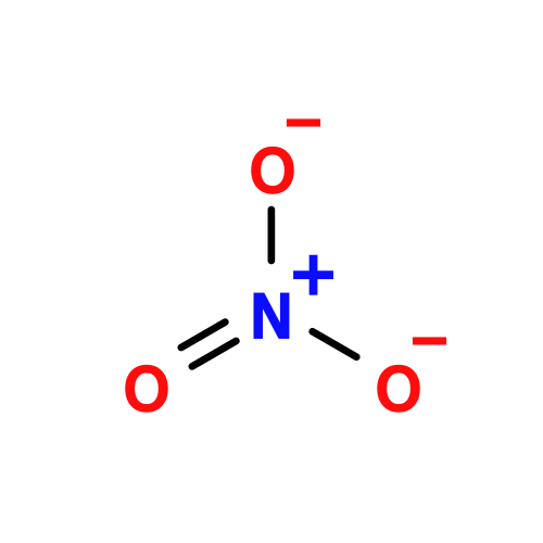 Mass: 62.005 Da / Num. of mol.: 1 / Source method: obtained synthetically / Formula: NO3 Mass: 62.005 Da / Num. of mol.: 1 / Source method: obtained synthetically / Formula: NO3#4: Water | ChemComp-HOH / |  Mass: 18.015 Da / Num. of mol.: 334 / Source method: isolated from a natural source / Formula: H2O Mass: 18.015 Da / Num. of mol.: 334 / Source method: isolated from a natural source / Formula: H2OHas ligand of interest | N | Has protein modification | Y | |
-Experimental details
-Experiment
| Experiment | Method: ELECTRON CRYSTALLOGRAPHY |
|---|---|
| EM experiment | Aggregation state: 3D ARRAY / 3D reconstruction method: electron crystallography |
- Sample preparation
| Component | Name: Proteinase K / Type: COMPLEX / Details: Serine protease / Entity ID: #1 / Source: RECOMBINANT |
|---|---|
| Molecular weight | Value: 0.0289 MDa / Experimental value: NO |
| Source (natural) | Organism: Parengyodontium album (fungus) |
| Source (recombinant) | Organism: Parengyodontium album (fungus) |
| Buffer solution | pH: 6.5 |
| Specimen | Conc.: 40 mg/ml / Embedding applied: NO / Shadowing applied: NO / Staining applied: NO / Vitrification applied: YES / Details: Microcrystals |
| Specimen support | Grid material: COPPER / Grid mesh size: 200 divisions/in. / Grid type: Quantifoil R2/2 |
| Vitrification | Instrument: LEICA PLUNGER / Cryogen name: ETHANE / Humidity: 95 % / Chamber temperature: 277 K |
-Data collection
| Experimental equipment |  Model: Titan Krios / Image courtesy: FEI Company |
|---|---|
| Microscopy | Model: TFS KRIOS |
| Electron gun | Electron source:  FIELD EMISSION GUN / Accelerating voltage: 300 kV / Illumination mode: FLOOD BEAM FIELD EMISSION GUN / Accelerating voltage: 300 kV / Illumination mode: FLOOD BEAM |
| Electron lens | Mode: DIFFRACTION / Nominal defocus max: 0 nm / Nominal defocus min: 0 nm / C2 aperture diameter: 50 µm / Alignment procedure: BASIC |
| Specimen holder | Cryogen: NITROGEN / Specimen holder model: FEI TITAN KRIOS AUTOGRID HOLDER / Temperature (max): 90 K / Temperature (min): 77 K |
| Image recording | Average exposure time: 1 sec. / Electron dose: 0.002 e/Å2 / Film or detector model: FEI FALCON IV (4k x 4k) / Num. of diffraction images: 420 / Num. of grids imaged: 1 / Num. of real images: 1 |
| EM imaging optics | Energyfilter name: TFS Selectris / Energyfilter slit width: 20 eV |
| Image scans | Sampling size: 14 µm / Width: 4096 / Height: 4096 |
| EM diffraction | Camera length: 1402 mm |
| EM diffraction shell | Resolution: 1.2→47.32 Å / Fourier space coverage: 97.9 % / Multiplicity: 13.3 / Num. of structure factors: 75213 / Phase residual: 14.3 ° |
| EM diffraction stats | Fourier space coverage: 97.9 % / High resolution: 1.2 Å / Num. of intensities measured: 1000823 / Num. of structure factors: 75213 / Phase error rejection criteria: None / Rmerge: 25.1 |
| Reflection | Biso Wilson estimate: 8.43 Å2 |
- Processing
| EM software |
| |||||||||||||||||||||||||||||||||||||||||||||||||||||||||||||||||||||||||||||||||||||||||||||||||||||||||||||||||||||||||||||||||||||||||||||||||||||||||||||||||||||||||||||||
|---|---|---|---|---|---|---|---|---|---|---|---|---|---|---|---|---|---|---|---|---|---|---|---|---|---|---|---|---|---|---|---|---|---|---|---|---|---|---|---|---|---|---|---|---|---|---|---|---|---|---|---|---|---|---|---|---|---|---|---|---|---|---|---|---|---|---|---|---|---|---|---|---|---|---|---|---|---|---|---|---|---|---|---|---|---|---|---|---|---|---|---|---|---|---|---|---|---|---|---|---|---|---|---|---|---|---|---|---|---|---|---|---|---|---|---|---|---|---|---|---|---|---|---|---|---|---|---|---|---|---|---|---|---|---|---|---|---|---|---|---|---|---|---|---|---|---|---|---|---|---|---|---|---|---|---|---|---|---|---|---|---|---|---|---|---|---|---|---|---|---|---|---|---|---|---|---|
| EM 3D crystal entity | ∠α: 90 ° / ∠β: 90 ° / ∠γ: 90 ° / A: 67.41 Å / B: 67.41 Å / C: 105.78 Å / Space group name: P43212 / Space group num: 96 | |||||||||||||||||||||||||||||||||||||||||||||||||||||||||||||||||||||||||||||||||||||||||||||||||||||||||||||||||||||||||||||||||||||||||||||||||||||||||||||||||||||||||||||||
| CTF correction | Type: NONE | |||||||||||||||||||||||||||||||||||||||||||||||||||||||||||||||||||||||||||||||||||||||||||||||||||||||||||||||||||||||||||||||||||||||||||||||||||||||||||||||||||||||||||||||
| 3D reconstruction | Resolution: 1.2 Å / Resolution method: DIFFRACTION PATTERN/LAYERLINES / Symmetry type: 3D CRYSTAL | |||||||||||||||||||||||||||||||||||||||||||||||||||||||||||||||||||||||||||||||||||||||||||||||||||||||||||||||||||||||||||||||||||||||||||||||||||||||||||||||||||||||||||||||
| Atomic model building | B value: 10.74 / Protocol: OTHER / Space: RECIPROCAL / Target criteria: Maximum likelihood | |||||||||||||||||||||||||||||||||||||||||||||||||||||||||||||||||||||||||||||||||||||||||||||||||||||||||||||||||||||||||||||||||||||||||||||||||||||||||||||||||||||||||||||||
| Atomic model building | PDB-ID: 9dho Accession code: 9dho / Details: Molecular replacement / Source name: PDB / Type: experimental model | |||||||||||||||||||||||||||||||||||||||||||||||||||||||||||||||||||||||||||||||||||||||||||||||||||||||||||||||||||||||||||||||||||||||||||||||||||||||||||||||||||||||||||||||
| Refinement | Resolution: 1.2→47.67 Å / SU ML: 0.1447 / Cross valid method: FREE R-VALUE / σ(F): 1.33 / Phase error: 19.0895 Stereochemistry target values: GeoStd + Monomer Library + CDL v1.2
| |||||||||||||||||||||||||||||||||||||||||||||||||||||||||||||||||||||||||||||||||||||||||||||||||||||||||||||||||||||||||||||||||||||||||||||||||||||||||||||||||||||||||||||||
| Solvent computation | Shrinkage radii: 0.9 Å / VDW probe radii: 1.1 Å / Solvent model: FLAT BULK SOLVENT MODEL | |||||||||||||||||||||||||||||||||||||||||||||||||||||||||||||||||||||||||||||||||||||||||||||||||||||||||||||||||||||||||||||||||||||||||||||||||||||||||||||||||||||||||||||||
| Displacement parameters | Biso mean: 10.74 Å2 | |||||||||||||||||||||||||||||||||||||||||||||||||||||||||||||||||||||||||||||||||||||||||||||||||||||||||||||||||||||||||||||||||||||||||||||||||||||||||||||||||||||||||||||||
| Refine LS restraints |
| |||||||||||||||||||||||||||||||||||||||||||||||||||||||||||||||||||||||||||||||||||||||||||||||||||||||||||||||||||||||||||||||||||||||||||||||||||||||||||||||||||||||||||||||
| LS refinement shell |
|