 Movie
Movie Controller

[English] 日本語
 Yorodumi
- PDB-9mue: Cryo-EM structure of CRISPR-associated cA4 bound Cat1 Pentagonal ... -
+ Open data

- Basic information
| Entry | Database: PDB / ID: 9mue | |||||||||
|---|---|---|---|---|---|---|---|---|---|---|
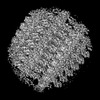
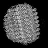
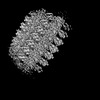
| Title | Cryo-EM structure of CRISPR-associated cA4 bound Cat1 Pentagonal filament assembly in the presence of NAD (ADPR modelled) | |||||||||
 Components Components |
| |||||||||
 Keywords Keywords | ANTIVIRAL PROTEIN / CRISPR / antiphage defense / adaptive immunity / Filament / CARF / TIR / NAD / ADPR / NAM | |||||||||
| Function / homology | Chem-AR6 / RNA Function and homology information Function and homology information | |||||||||
| Biological species |  bacterium (bacteria) bacterium (bacteria) | |||||||||
| Method | ELECTRON MICROSCOPY / single particle reconstruction / cryo EM / Resolution: 4 Å | |||||||||
 Authors Authors | Majumder, P. / Patel, D.J. | |||||||||
| Funding support |  United States, 2items United States, 2items
| |||||||||
 Citation Citation | Journal: Science / Year: 2025 Title: Cat1 forms filament networks to degrade NAD during the type III CRISPR-Cas antiviral response. Authors: Christian F Baca / Puja Majumder / James H Hickling / Dinshaw J Patel / Luciano A Marraffini / Abstract: Type III CRISPR-Cas systems defend against viral infection in prokaryotes by using an RNA-guided complex that recognizes foreign transcripts and synthesizes cyclic oligoadenylate (cOA) messengers to ...Type III CRISPR-Cas systems defend against viral infection in prokaryotes by using an RNA-guided complex that recognizes foreign transcripts and synthesizes cyclic oligoadenylate (cOA) messengers to activate CRISPR-associated Rossmann-fold (CARF) immune effectors. In this study, we investigated a protein containing a CARF domain-fused Toll/interleukin-1 receptor (TIR) domain, Cat1. We found that Cat1 provides immunity by cleaving and depleting oxidized nicotinamide adenine dinucleotide (NAD) molecules from the infected host, inducing a growth arrest that prevents viral propagation. Cat1 forms dimers that stack upon each other to generate long filaments that are maintained by bound cOA ligands, with stacked TIR domains forming the NAD cleavage catalytic sites. Furthermore, Cat1 filaments assemble into distinct trigonal and pentagonal networks that enhance NAD degradation. Cat1 presents an unprecedented chemistry and higher-order protein assembly for the CRISPR-Cas response. | |||||||||
| History |
|
- Structure visualization
| Structure viewer | Molecule: MolmilJmol/JSmol |
|---|
- Downloads & links
-Download
| PDBx/mmCIF format | 9mue.cif.gz | 190.8 KB | Display | PDBx/mmCIF format |
|---|---|---|---|---|
| PDB format | pdb9mue.ent.gz | 154.1 KB | Display | PDB format |
| PDBx/mmJSON format | 9mue.json.gz | Tree view | PDBx/mmJSON format | |
| Others |  Other downloads Other downloads |
-Validation report
| Arichive directory | https://data.pdbj.org/pub/pdb/validation_reports/mu/9mueftp://data.pdbj.org/pub/pdb/validation_reports/mu/9mue | HTTPS FTP |
|---|
-Related structure data
| Related structure data | 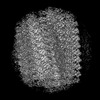 48630MC 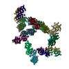 9mudC 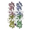 9muoC  9mw9C M: map data used to model this data C: citing same article ( |
|---|---|
| Similar structure data |
-Links
 PDBj
PDBj

- Assembly
| Deposited unit | 
|
|---|---|
| 1 |
|
-Components
| #1: Protein | Mass: 30150.592 Da / Num. of mol.: 4 Source method: isolated from a genetically manipulated source Source: (gene. exp.) bacterium (bacteria) / Production host: #2: RNA chain | Mass: 1271.866 Da / Num. of mol.: 2 / Source method: isolated from a natural source / Source: (natural) bacterium (bacteria)#3: Chemical |   Mass: 559.316 Da / Num. of mol.: 2 / Source method: obtained synthetically / Formula: C15H23N5O14P2 / Feature type: SUBJECT OF INVESTIGATION Mass: 559.316 Da / Num. of mol.: 2 / Source method: obtained synthetically / Formula: C15H23N5O14P2 / Feature type: SUBJECT OF INVESTIGATIONHas ligand of interest | Y | Has protein modification | N | |
|---|
-Experimental details
-Experiment
| Experiment | Method: ELECTRON MICROSCOPY |
|---|---|
| EM experiment | Aggregation state: FILAMENT / 3D reconstruction method: single particle reconstruction |
- Sample preparation
| Component | Name: Antiviral protein Cat1 - Cyclic Tetra-Adenylate Complex NAD substrate was mixed with the sample. Type: COMPLEX Details: Cat1 protein forms filament structure upon recognition of cA4, Cyclic Tetra-Adenylate ligand. NAD substrate was mixed with the sample. Entity ID: #1-#2 / Source: RECOMBINANT |
|---|---|
| Source (natural) | Organism: bacterium (bacteria) |
| Source (recombinant) | Organism: |
| Buffer solution | pH: 6 |
| Specimen | Conc.: 1.2 mg/ml / Embedding applied: NO / Shadowing applied: NO / Staining applied: NO / Vitrification applied: YES |
| Specimen support | Grid material: GOLD / Grid mesh size: 300 divisions/in. / Grid type: Quantifoil R1.2/1.3 |
| Vitrification | Instrument: FEI VITROBOT MARK IV / Cryogen name: ETHANE / Humidity: 100 % / Chamber temperature: 277 K Details: liquid-nitrogen-cooled liquid ethane was used as the cryogen. |
- Electron microscopy imaging
| Experimental equipment |  Model: Titan Krios / Image courtesy: FEI Company |
|---|---|
| Microscopy | Model: TFS KRIOS |
| Electron gun | Electron source:  FIELD EMISSION GUN / Accelerating voltage: 300 kV / Illumination mode: FLOOD BEAM FIELD EMISSION GUN / Accelerating voltage: 300 kV / Illumination mode: FLOOD BEAM |
| Electron lens | Mode: BRIGHT FIELD / Nominal defocus max: 2000 nm / Nominal defocus min: 800 nm / Cs: 2.7 mm |
| Specimen holder | Cryogen: NITROGEN |
| Image recording | Electron dose: 57.32 e/Å2 / Film or detector model: GATAN K3 (6k x 4k) |
- Processing
| EM software |
| ||||||||||||||||||||||||
|---|---|---|---|---|---|---|---|---|---|---|---|---|---|---|---|---|---|---|---|---|---|---|---|---|---|
| CTF correction | Type: NONE | ||||||||||||||||||||||||
| 3D reconstruction | Resolution: 4 Å / Resolution method: FSC 0.143 CUT-OFF / Num. of particles: 152750 / Symmetry type: POINT | ||||||||||||||||||||||||
| Refine LS restraints |
|
