[English] 日本語
Yorodumi
- PDB-9mhb: Crystal Structure of Human P38 alpha MAPK In Complex with MW01-14... -

+
Open data


ID or keywords:

Loading...

-
Basic information

Entry
Database: PDB / ID: 9mhb
TitleCrystal Structure of Human P38 alpha MAPK In Complex with MW01-14-064SRM
ComponentsMitogen-activated protein kinase 14
KeywordsTRANSFERASE/INHIBITOR / P38 alpha MAPK / MW01-14-064SRM / TRANSFERASE / TRANSFERASE-INHIBITOR complex
Function / homology
Function and homology information


stress-activated protein kinase signaling cascade / positive regulation of cyclase activity / Activation of PPARGC1A (PGC-1alpha) by phosphorylation / regulation of synaptic membrane adhesion / stress-induced premature senescence / CD163 mediating an anti-inflammatory response / cell surface receptor protein serine/threonine kinase signaling pathway / 3'-UTR-mediated mRNA stabilization / positive regulation of myoblast fusion / KSRP (KHSRP) binds and destabilizes mRNA ...stress-activated protein kinase signaling cascade / positive regulation of cyclase activity / Activation of PPARGC1A (PGC-1alpha) by phosphorylation / regulation of synaptic membrane adhesion / stress-induced premature senescence / CD163 mediating an anti-inflammatory response / cell surface receptor protein serine/threonine kinase signaling pathway / 3'-UTR-mediated mRNA stabilization / positive regulation of myoblast fusion / KSRP (KHSRP) binds and destabilizes mRNA / cartilage condensation / cellular response to UV-B / mitogen-activated protein kinase p38 binding / Platelet sensitization by LDL / positive regulation of muscle cell differentiation / Myogenesis / positive regulation of myotube differentiation / NFAT protein binding / regulation of cytokine production involved in inflammatory response / Activation of the AP-1 family of transcription factors / D-glucose import / p38MAPK cascade / ERK/MAPK targets / Regulation of MITF-M-dependent genes involved in pigmentation / fatty acid oxidation / cellular response to lipoteichoic acid / response to dietary excess / response to muramyl dipeptide / MAP kinase kinase activity / JUN kinase activity / RHO GTPases Activate NADPH Oxidases / regulation of ossification / MAP kinase activity / cellular response to vascular endothelial growth factor stimulus / signal transduction in response to DNA damage / mitogen-activated protein kinase / chondrocyte differentiation / negative regulation of hippo signaling / positive regulation of myoblast differentiation / vascular endothelial growth factor receptor signaling pathway / skeletal muscle tissue development / stress-activated MAPK cascade / positive regulation of cardiac muscle cell proliferation / p38MAPK events / striated muscle cell differentiation / response to muscle stretch / positive regulation of brown fat cell differentiation / positive regulation of interleukin-12 production / osteoclast differentiation / lipopolysaccharide-mediated signaling pathway / positive regulation of erythrocyte differentiation / DNA damage checkpoint signaling / cellular response to ionizing radiation / positive regulation of D-glucose import / response to insulin / activated TAK1 mediates p38 MAPK activation / stem cell differentiation / placenta development / negative regulation of inflammatory response to antigenic stimulus / negative regulation of canonical Wnt signaling pathway / NOD1/2 Signaling Pathway / cell morphogenesis / bone development / platelet activation / cellular response to virus / positive regulation of protein import into nucleus / VEGFA-VEGFR2 Pathway / glucose metabolic process / positive regulation of reactive oxygen species metabolic process / spindle pole / chemotaxis / osteoblast differentiation / ADP signalling through P2Y purinoceptor 1 / cellular senescence / peptidyl-serine phosphorylation / cellular response to tumor necrosis factor / cellular response to lipopolysaccharide / secretory granule lumen / protein phosphatase binding / Oxidative Stress Induced Senescence / angiogenesis / Regulation of TP53 Activity through Phosphorylation / ficolin-1-rich granule lumen / transcription by RNA polymerase II / cell surface receptor signaling pathway / nuclear speck / intracellular signal transduction / protein serine kinase activity / protein serine/threonine kinase activity / apoptotic process / Neutrophil degranulation / positive regulation of gene expression / regulation of transcription by RNA polymerase II / glutamatergic synapse / enzyme binding / signal transduction / positive regulation of transcription by RNA polymerase II / mitochondrion / extracellular region / nucleoplasm
Similarity search - Function
Mitogen-activated protein kinase 14 / Mitogen-activated protein (MAP) kinase p38-like / Mitogen-activated protein (MAP) kinase, conserved site / MAP kinase signature. / : / Protein kinase domain / Serine/Threonine protein kinases, catalytic domain / Protein kinase, ATP binding site / Protein kinases ATP-binding region signature. / Protein kinase domain profile. ...Mitogen-activated protein kinase 14 / Mitogen-activated protein (MAP) kinase p38-like / Mitogen-activated protein (MAP) kinase, conserved site / MAP kinase signature. / : / Protein kinase domain / Serine/Threonine protein kinases, catalytic domain / Protein kinase, ATP binding site / Protein kinases ATP-binding region signature. / Protein kinase domain profile. / Protein kinase domain / Protein kinase-like domain superfamily
Similarity search - Domain/homology
: / ACETATE ION / 4-[3-(4-FLUOROPHENYL)-1H-PYRAZOL-4-YL]PYRIDINE / DI(HYDROXYETHYL)ETHER / Mitogen-activated protein kinase 14
Similarity search - Component
Biological speciesHomo sapiens (human)
MethodX-RAY DIFFRACTION / SYNCHROTRON / MOLECULAR REPLACEMENT / Resolution: 1.65 Å
AuthorsMinasov, G. / Tokars, V.L. / Roy, S.M. / Watterson, D.M. / Shuvalova, L.
Funding support United States, 1items
OrganizationGrant numberCountry
National Institutes of Health/National Institute on Aging (NIH/NIA)AG066722 United States
CitationJournal: To Be Published
Title: Crystal Structure of Human P38 alpha MAPK In Complex with MW01-14-064SRM
Authors: Minasov, G. / Tokars, V.L. / Roy, S.M. / Watterson, D.M. / Shuvalova, L.
History
DepositionDec 11, 2024Deposition site: RCSB / Processing site: RCSB
Revision 1.0Jan 1, 2025Provider: repository / Type: Initial release

-
Structure visualization

Structure viewerMolecule:
MolmilJmol/JSmol

Downloads & links

-
Assembly

Deposited unit
A: Mitogen-activated protein kinase 14
hetero molecules


Theoretical massNumber of molelcules
Total (without water)42,6528
Polymers41,5601
Non-polymers1,0917
Water5,819323
1


  • Idetical with deposited unit
  • defined by author
TypeNameSymmetry operationNumber
identity operation1_555x,y,z1
Unit cell
Length a, b, c (Å)66.446, 74.862, 78.065
Angle α, β, γ (deg.)90.00, 90.00, 90.00
Int Tables number19
Space group name H-MP212121

-
Components

-
Protein , 1 types, 1 molecules A

#1: Protein Mitogen-activated protein kinase 14 / MAP kinase 14 / MAPK 14 / Cytokine suppressive anti-inflammatory drug-binding protein / CSAID- ...MAP kinase 14 / MAPK 14 / Cytokine suppressive anti-inflammatory drug-binding protein / CSAID-binding protein / CSBP / MAP kinase MXI2 / MAX-interacting protein 2 / Mitogen-activated protein kinase p38 alpha / MAP kinase p38 alpha / Stress-activated protein kinase 2a / SAPK2a


Mass: 41560.375 Da / Num. of mol.: 1
Source method: isolated from a genetically manipulated source
Source: (gene. exp.) Homo sapiens (human) / Gene: MAPK14, CSBP, CSBP1, CSBP2, CSPB1, MXI2, SAPK2A / Plasmid: pMCSG7 / Production host: Escherichia coli BL21(DE3) (bacteria) / Variant (production host): pLysS
References: UniProt: Q16539, mitogen-activated protein kinase

-
Non-polymers , 6 types, 330 molecules

#2: Chemical ChemComp-A1BLA / (5P)-N,N-dimethyl-5-(naphthalen-1-yl)-6-(pyridin-4-yl)pyrazin-2-amine


Mass: 326.394 Da / Num. of mol.: 1 / Source method: obtained synthetically / Formula: C21H18N4 / Feature type: SUBJECT OF INVESTIGATION
#3: Chemical ChemComp-GG5 / 4-[3-(4-FLUOROPHENYL)-1H-PYRAZOL-4-YL]PYRIDINE


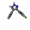
Mass: 239.248 Da / Num. of mol.: 2 / Source method: obtained synthetically / Formula: C14H10FN3 / Feature type: SUBJECT OF INVESTIGATION
#4: Chemical ChemComp-ACT / ACETATE ION


Mass: 59.044 Da / Num. of mol.: 2 / Source method: obtained synthetically / Formula: C2H3O2
#5: Chemical ChemComp-PEG / DI(HYDROXYETHYL)ETHER


Mass: 106.120 Da / Num. of mol.: 1 / Source method: obtained synthetically / Formula: C4H10O3
#6: Chemical ChemComp-EDO / 1,2-ETHANEDIOL / ETHYLENE GLYCOL


Mass: 62.068 Da / Num. of mol.: 1 / Source method: obtained synthetically / Formula: C2H6O2
#7: Water ChemComp-HOH / water


Mass: 18.015 Da / Num. of mol.: 323 / Source method: isolated from a natural source / Formula: H2O

-
Details

Has ligand of interestY
Has protein modificationY

-
Experimental details

-
Experiment

ExperimentMethod: X-RAY DIFFRACTION / Number of used crystals: 1

-
Sample preparation

CrystalDensity Matthews: 2.4 Å3/Da / Density % sol: 48.6 %
Crystal growTemperature: 292 K / Method: vapor diffusion, sitting drop / pH: 5.5
Details: Protein: 20.0 mg/ml, 0.15M Sodium chloride, 0.02M Tris-HCl (pH 8.3), 1mM TCEP; Screen: 0.15M Ammomium acetate, 0.1M Bis-Tris (pH 5.5), 16% (w/v) PEG1000; Soak: 3 hrs, 5mM ligand in screen ...Details: Protein: 20.0 mg/ml, 0.15M Sodium chloride, 0.02M Tris-HCl (pH 8.3), 1mM TCEP; Screen: 0.15M Ammomium acetate, 0.1M Bis-Tris (pH 5.5), 16% (w/v) PEG1000; Soak: 3 hrs, 5mM ligand in screen solution; Cryo: 10% Glycerol in screen solution.

-
Data collection

DiffractionMean temperature: 100 K / Serial crystal experiment: N
Diffraction sourceSource: SYNCHROTRON / Site: APS / Beamline: 21-ID-G / Wavelength: 0.97856 Å
DetectorType: MARMOSAIC 300 mm CCD / Detector: CCD / Date: Oct 10, 2011
RadiationMonochromator: C(111) / Protocol: SINGLE WAVELENGTH / Monochromatic (M) / Laue (L): M / Scattering type: x-ray
Radiation wavelengthWavelength: 0.97856 Å / Relative weight: 1
ReflectionResolution: 1.65→30 Å / Num. obs: 47571 / % possible obs: 100 % / Observed criterion σ(I): -3 / Redundancy: 7.4 % / Biso Wilson estimate: 26 Å2 / CC1/2: 0.997 / CC star: 0.999 / Rmerge(I) obs: 0.044 / Rpim(I) all: 0.018 / Rrim(I) all: 0.048 / Rsym value: 0.044 / Χ2: 1.005 / Net I/σ(I): 44.9
Reflection shellResolution: 1.65→1.68 Å / Redundancy: 7.4 % / Rmerge(I) obs: 0.969 / Mean I/σ(I) obs: 2.3 / Num. unique obs: 2343 / CC1/2: 0.746 / CC star: 0.924 / Rpim(I) all: 0.381 / Rsym value: 0.969 / Χ2: 1.002 / % possible all: 100

-
Processing

Software
NameVersionClassification
REFMAC5.8.0425refinement
HKL-3000data reduction
HKL-3000data scaling
PHASERphasing
RefinementMethod to determine structure: MOLECULAR REPLACEMENT / Resolution: 1.65→28.3 Å / Cor.coef. Fo:Fc: 0.963 / Cor.coef. Fo:Fc free: 0.949 / SU B: 3.618 / SU ML: 0.063 / Cross valid method: THROUGHOUT / ESU R: 0.093 / ESU R Free: 0.091 / Stereochemistry target values: MAXIMUM LIKELIHOOD / Details: HYDROGENS HAVE BEEN ADDED IN THE RIDING POSITIONS
RfactorNum. reflection% reflectionSelection details
Rfree0.20174 2273 4.8 %RANDOM
Rwork0.17254 ---
obs0.17393 45229 99.92 %-
Solvent computationIon probe radii: 0.8 Å / Shrinkage radii: 0.8 Å / VDW probe radii: 1.2 Å / Solvent model: MASK
Displacement parametersBiso mean: 31.536 Å2
Baniso -1Baniso -2Baniso -3
1-0.2 Å2-0 Å2-0 Å2
2---0.52 Å20 Å2
3---0.33 Å2
Refinement stepCycle: 1 / Resolution: 1.65→28.3 Å
ProteinNucleic acidLigandSolventTotal
Num. atoms2724 0 84 323 3131
Refine LS restraints
Refine-IDTypeDev idealDev ideal targetNumber
X-RAY DIFFRACTIONr_bond_refined_d0.0050.0123035
X-RAY DIFFRACTIONr_bond_other_d0.0010.0162885
X-RAY DIFFRACTIONr_angle_refined_deg1.5241.8444133
X-RAY DIFFRACTIONr_angle_other_deg0.5191.7766641
X-RAY DIFFRACTIONr_dihedral_angle_1_deg3.0335367
X-RAY DIFFRACTIONr_dihedral_angle_2_deg4.462524
X-RAY DIFFRACTIONr_dihedral_angle_3_deg7.39110525
X-RAY DIFFRACTIONr_dihedral_angle_4_deg
X-RAY DIFFRACTIONr_chiral_restr0.080.2449
X-RAY DIFFRACTIONr_gen_planes_refined0.0170.023603
X-RAY DIFFRACTIONr_gen_planes_other0.0130.02714
X-RAY DIFFRACTIONr_nbd_refined
X-RAY DIFFRACTIONr_nbd_other
X-RAY DIFFRACTIONr_nbtor_refined
X-RAY DIFFRACTIONr_nbtor_other
X-RAY DIFFRACTIONr_xyhbond_nbd_refined
X-RAY DIFFRACTIONr_xyhbond_nbd_other
X-RAY DIFFRACTIONr_metal_ion_refined
X-RAY DIFFRACTIONr_metal_ion_other
X-RAY DIFFRACTIONr_symmetry_vdw_refined
X-RAY DIFFRACTIONr_symmetry_vdw_other
X-RAY DIFFRACTIONr_symmetry_hbond_refined
X-RAY DIFFRACTIONr_symmetry_hbond_other
X-RAY DIFFRACTIONr_symmetry_metal_ion_refined
X-RAY DIFFRACTIONr_symmetry_metal_ion_other
X-RAY DIFFRACTIONr_mcbond_it2.0851.7921428
X-RAY DIFFRACTIONr_mcbond_other2.0511.7871426
X-RAY DIFFRACTIONr_mcangle_it3.1773.1931804
X-RAY DIFFRACTIONr_mcangle_other3.1763.1941805
X-RAY DIFFRACTIONr_scbond_it3.1312.2231607
X-RAY DIFFRACTIONr_scbond_other3.132.2251608
X-RAY DIFFRACTIONr_scangle_it
X-RAY DIFFRACTIONr_scangle_other4.8063.92329
X-RAY DIFFRACTIONr_long_range_B_refined8.09322.993622
X-RAY DIFFRACTIONr_long_range_B_other8.00921.623539
X-RAY DIFFRACTIONr_rigid_bond_restr
X-RAY DIFFRACTIONr_sphericity_free
X-RAY DIFFRACTIONr_sphericity_bonded
LS refinement shellResolution: 1.65→1.693 Å / Total num. of bins used: 20
RfactorNum. reflection% reflection
Rfree0.249 161 -
Rwork0.239 3301 -
obs--99.57 %
Refinement TLS params.

Method: refined / Refine-ID: X-RAY DIFFRACTION

IDL112)L122)L132)L222)L232)L332)S11 (Å °)S12 (Å °)S13 (Å °)S21 (Å °)S22 (Å °)S23 (Å °)S31 (Å °)S32 (Å °)S33 (Å °)T112)T122)T132)T222)T232)T332)Origin x (Å)Origin y (Å)Origin z (Å)
13.64690.70020.66846.9171-2.23523.8711-0.0266-0.3024-0.10270.70020.18130.5488-0.3745-0.2768-0.15480.15620.00430.06490.11470.10260.1519-16.473-0.22221.157
21.9045-1.01670.383.1754-1.77281.64490.0752-0.0455-0.10160.14660.06870.1937-0.0941-0.1298-0.14390.1018-0.0007-0.00510.01590.01090.0448-7.4665.01511.193
35.8178-1.53251.63440.5276-0.66631.43720.0686-0.63230.04020.08160.12140.1211-0.2108-0.1568-0.190.2917-0.06310.04050.20880.03240.2234-3.3660.6421.537
42.15490.74030.09792.3934-0.7031.40050.0942-0.0862-0.11260.0596-0.15460.09380.12720.08630.06040.1123-0.00420.01350.04360.03750.06810.708-3.78118.213
52.35531.0539-0.2562.7471-0.47081.44260.1038-0.18970.17710.2149-0.1084-0.0346-0.02730.08260.00460.0792-0.017-0.01490.06540.0330.064320.79510.89119.3
61.6339-0.008-0.08171.5419-0.20080.43170.0239-0.1714-0.12910.0827-0.1282-0.42710.0320.18820.10420.13790.0205-0.00270.13930.07430.139924.7291.90715.503
72.5178-1.44680.40095.7816-4.36483.62540.07760.1715-0.1297-0.3830.01840.29090.2541-0.0547-0.0960.15280.0303-0.04290.07890.01370.1192-8.4897.5812.587
Refinement TLS group
IDRefine-IDRefine TLS-IDAuth asym-IDAuth seq-ID
1X-RAY DIFFRACTION1A4 - 48
2X-RAY DIFFRACTION2A49 - 96
3X-RAY DIFFRACTION3A97 - 120
4X-RAY DIFFRACTION4A121 - 167
5X-RAY DIFFRACTION5A168 - 250
6X-RAY DIFFRACTION6A251 - 324
7X-RAY DIFFRACTION7A325 - 353

+
About Yorodumi

-
News

-
Feb 9, 2022. New format data for meta-information of EMDB entries

New format data for meta-information of EMDB entries

  • Version 3 of the EMDB header file is now the official format.
  • The previous official version 1.9 will be removed from the archive.

Related info.:EMDB header

External links:wwPDB to switch to version 3 of the EMDB data model

-
Aug 12, 2020. Covid-19 info

Covid-19 info

URL: https://pdbj.org/emnavi/covid19.php

New page: Covid-19 featured information page in EM Navigator.

Related info.:Covid-19 info / Mar 5, 2020. Novel coronavirus structure data

+
Mar 5, 2020. Novel coronavirus structure data

Novel coronavirus structure data

Related info.:Yorodumi Speices / Aug 12, 2020. Covid-19 info

External links:COVID-19 featured content - PDBj / Molecule of the Month (242):Coronavirus Proteases

+
Jan 31, 2019. EMDB accession codes are about to change! (news from PDBe EMDB page)

EMDB accession codes are about to change! (news from PDBe EMDB page)

  • The allocation of 4 digits for EMDB accession codes will soon come to an end. Whilst these codes will remain in use, new EMDB accession codes will include an additional digit and will expand incrementally as the available range of codes is exhausted. The current 4-digit format prefixed with “EMD-” (i.e. EMD-XXXX) will advance to a 5-digit format (i.e. EMD-XXXXX), and so on. It is currently estimated that the 4-digit codes will be depleted around Spring 2019, at which point the 5-digit format will come into force.
  • The EM Navigator/Yorodumi systems omit the EMD- prefix.

Related info.:Q: What is EMD? / ID/Accession-code notation in Yorodumi/EM Navigator

External links:EMDB Accession Codes are Changing Soon! / Contact to PDBj

+
Jul 12, 2017. Major update of PDB

Major update of PDB

  • wwPDB released updated PDB data conforming to the new PDBx/mmCIF dictionary.
  • This is a major update changing the version number from 4 to 5, and with Remediation, in which all the entries are updated.
  • In this update, many items about electron microscopy experimental information are reorganized (e.g. em_software).
  • Now, EM Navigator and Yorodumi are based on the updated data.

External links:wwPDB Remediation / Enriched Model Files Conforming to OneDep Data Standards Now Available in the PDB FTP Archive

-
Yorodumi

Thousand views of thousand structures

  • Yorodumi is a browser for structure data from EMDB, PDB, SASBDB, etc.
  • This page is also the successor to EM Navigator detail page, and also detail information page/front-end page for Omokage search.
  • The word "yorodu" (or yorozu) is an old Japanese word meaning "ten thousand". "mi" (miru) is to see.

Related info.:EMDB / PDB / SASBDB / Comparison of 3 databanks / Yorodumi Search / Aug 31, 2016. New EM Navigator & Yorodumi / Yorodumi Papers / Jmol/JSmol / Function and homology information / Changes in new EM Navigator and Yorodumi

Read more