 Movie
Movie Controller

[English] 日本語
 Yorodumi
- PDB-9mgs: Structure of aminoglycoside acetyltransferase AAC(3)-Ia in comple... -
+ Open data

- Basic information
| Entry | Database: PDB / ID: 9mgs | ||||||
|---|---|---|---|---|---|---|---|
| Title | Structure of aminoglycoside acetyltransferase AAC(3)-Ia in complex with sisomicin and CoA | ||||||
 Components Components | Aminoglycoside acetyltransferase | ||||||
 Keywords Keywords | TRANSFERASE / antibiotic resistance / aminoglycosides | ||||||
| Function / homology | acyltransferase activity, transferring groups other than amino-acyl groups / Acetyltransferase (GNAT) family / Gcn5-related N-acetyltransferase (GNAT) domain profile. / GNAT domain / Acyl-CoA N-acyltransferase / COENZYME A / Chem-SIS / Aminoglycoside acetyltransferase Function and homology information Function and homology information | ||||||
| Biological species |  Acinetobacter baumannii (bacteria) Acinetobacter baumannii (bacteria) | ||||||
| Method |  X-RAY DIFFRACTION / SYNCHROTRON / MOLECULAR REPLACEMENT / Resolution: 2.1 Å X-RAY DIFFRACTION / SYNCHROTRON / MOLECULAR REPLACEMENT / Resolution: 2.1 Å | ||||||
 Authors Authors | Hemmings, M. / Zielinski, M. / Blanchet, J. / Golkar, T. / Berghuis, A.M. | ||||||
| Funding support |  Canada, 1items Canada, 1items
| ||||||
 Citation Citation | Journal: Commun Chem / Year: 2025 Title: Enzyme-mediated aminoglycoside resistance without target mimicry. Authors: Hemmings, M. / Zielinski, M. / Golkar, T. / Blanchet, J. / Pistofidis, A. / Munro, K. / Schmeing, T.M. / Bohle, D.S. / Berghuis, A.M. | ||||||
| History |
|
- Structure visualization
| Structure viewer | Molecule: MolmilJmol/JSmol |
|---|
- Downloads & links
-Download
| PDBx/mmCIF format | 9mgs.cif.gz | 49.4 KB | Display | PDBx/mmCIF format |
|---|---|---|---|---|
| PDB format | pdb9mgs.ent.gz | 32.6 KB | Display | PDB format |
| PDBx/mmJSON format | 9mgs.json.gz | Tree view | PDBx/mmJSON format | |
| Others |  Other downloads Other downloads |
-Validation report
| Arichive directory | https://data.pdbj.org/pub/pdb/validation_reports/mg/9mgsftp://data.pdbj.org/pub/pdb/validation_reports/mg/9mgs | HTTPS FTP |
|---|
-Related structure data
| Related structure data |  9mgtC  9mguC  9mh3C  9mh5C  9mh6C  9mh7C C: citing same article ( |
|---|---|
| Similar structure data |
-Links
 PDBj
PDBj


- Assembly
| Deposited unit | 
| ||||||||
|---|---|---|---|---|---|---|---|---|---|
| 1 | 
| ||||||||
| Unit cell |
| ||||||||
| Components on special symmetry positions |
|
-Components
| #1: Protein | Mass: 16931.188 Da / Num. of mol.: 1 Source method: isolated from a genetically manipulated source Source: (gene. exp.) Acinetobacter baumannii (bacteria) / Gene: aac1Production host: References: UniProt: D7R512 |
|---|---|
| #2: Chemical | ChemComp-COA /   Mass: 767.534 Da / Num. of mol.: 1 / Source method: obtained synthetically / Formula: C21H36N7O16P3S / Feature type: SUBJECT OF INVESTIGATION Mass: 767.534 Da / Num. of mol.: 1 / Source method: obtained synthetically / Formula: C21H36N7O16P3S / Feature type: SUBJECT OF INVESTIGATION |
| #3: Chemical | ChemComp-SIS / (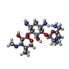  Mass: 447.526 Da / Num. of mol.: 1 / Source method: obtained synthetically / Formula: C19H37N5O7 / Feature type: SUBJECT OF INVESTIGATION / Comment: antibiotic*YM Mass: 447.526 Da / Num. of mol.: 1 / Source method: obtained synthetically / Formula: C19H37N5O7 / Feature type: SUBJECT OF INVESTIGATION / Comment: antibiotic*YM |
| #4: Water | ChemComp-HOH /  Mass: 18.015 Da / Num. of mol.: 61 / Source method: isolated from a natural source / Formula: H2O Mass: 18.015 Da / Num. of mol.: 61 / Source method: isolated from a natural source / Formula: H2O |
| Has ligand of interest | Y |
| Has protein modification | N |
-Experimental details
-Experiment
| Experiment | Method: X-RAY DIFFRACTION / Number of used crystals: 1 |
|---|
- Sample preparation
| Crystal | Density Matthews: 3.03 Å3/Da / Density % sol: 59.36 % |
|---|---|
| Crystal grow | Temperature: 293.15 K / Method: vapor diffusion, sitting drop Details: 10 mg/mL AAC(3)-Ia with a 10x molar excess of gentamicin and CoASH in 0.1 M Tris Ph 8.5, 0.05 M MgCl2, 20% EtOH, 35% PEG 400. |
-Data collection
| Diffraction | Mean temperature: 100 K / Serial crystal experiment: N |
|---|---|
| Diffraction source | Source: SYNCHROTRON / Site: CLSI / Beamline: 08B1-1 / Wavelength: 0.9537 Å |
| Detector | Type: DECTRIS PILATUS 6M / Detector: PIXEL / Date: Oct 1, 2022 |
| Radiation | Protocol: SINGLE WAVELENGTH / Monochromatic (M) / Laue (L): M / Scattering type: x-ray |
| Radiation wavelength | Wavelength: 0.9537 Å / Relative weight: 1 |
| Reflection | Resolution: 2.1→30 Å / Num. obs: 10939 / % possible obs: 85.07 % / Redundancy: 21.1 % / CC1/2: 0.992 / Net I/σ(I): 14.1 |
| Reflection shell | Resolution: 2.1→2.18 Å / Mean I/σ(I) obs: 1.2 / Num. unique obs: 1244 / CC1/2: 0.97 |
- Processing
| Software |
| ||||||||||||||||||||||||||||||||||||||||||||||||||||||||||||||||||||||||||||||||||||||||||||||||||||||||||||||||||||||||||||||||||||||||||||||||||||||||||||||||||||||||||||||||||||||
|---|---|---|---|---|---|---|---|---|---|---|---|---|---|---|---|---|---|---|---|---|---|---|---|---|---|---|---|---|---|---|---|---|---|---|---|---|---|---|---|---|---|---|---|---|---|---|---|---|---|---|---|---|---|---|---|---|---|---|---|---|---|---|---|---|---|---|---|---|---|---|---|---|---|---|---|---|---|---|---|---|---|---|---|---|---|---|---|---|---|---|---|---|---|---|---|---|---|---|---|---|---|---|---|---|---|---|---|---|---|---|---|---|---|---|---|---|---|---|---|---|---|---|---|---|---|---|---|---|---|---|---|---|---|---|---|---|---|---|---|---|---|---|---|---|---|---|---|---|---|---|---|---|---|---|---|---|---|---|---|---|---|---|---|---|---|---|---|---|---|---|---|---|---|---|---|---|---|---|---|---|---|---|---|
| Refinement | Method to determine structure: MOLECULAR REPLACEMENT / Resolution: 2.1→30 Å / Cor.coef. Fo:Fc: 0.937 / Cor.coef. Fo:Fc free: 0.915 / SU B: 7.172 / SU ML: 0.179 / Cross valid method: THROUGHOUT / ESU R: 0.259 / ESU R Free: 0.219 / Stereochemistry target values: MAXIMUM LIKELIHOOD / Details: HYDROGENS HAVE BEEN USED IF PRESENT IN THE INPUT
| ||||||||||||||||||||||||||||||||||||||||||||||||||||||||||||||||||||||||||||||||||||||||||||||||||||||||||||||||||||||||||||||||||||||||||||||||||||||||||||||||||||||||||||||||||||||
| Solvent computation | Ion probe radii: 0.8 Å / Shrinkage radii: 0.8 Å / VDW probe radii: 1.2 Å / Solvent model: MASK | ||||||||||||||||||||||||||||||||||||||||||||||||||||||||||||||||||||||||||||||||||||||||||||||||||||||||||||||||||||||||||||||||||||||||||||||||||||||||||||||||||||||||||||||||||||||
| Displacement parameters | Biso mean: 39.357 Å2
| ||||||||||||||||||||||||||||||||||||||||||||||||||||||||||||||||||||||||||||||||||||||||||||||||||||||||||||||||||||||||||||||||||||||||||||||||||||||||||||||||||||||||||||||||||||||
| Refinement step | Cycle: 1 / Resolution: 2.1→30 Å
| ||||||||||||||||||||||||||||||||||||||||||||||||||||||||||||||||||||||||||||||||||||||||||||||||||||||||||||||||||||||||||||||||||||||||||||||||||||||||||||||||||||||||||||||||||||||
| Refine LS restraints |
|