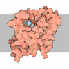
[English] 日本語
 Yorodumi
- PDB-9m8m: Structure of photosynthetic LH1-RC complex the Halophilic Nonsulf... -
+ Open data
- Basic information
| Entry | Database: PDB / ID: 9m8m | |||||||||||||||||||||
|---|---|---|---|---|---|---|---|---|---|---|---|---|---|---|---|---|---|---|---|---|---|---|
| Title | Structure of photosynthetic LH1-RC complex the Halophilic Nonsulfur Purple Bacterium, Rhodothalassium salexigens | |||||||||||||||||||||
 Components Components |
| |||||||||||||||||||||
 Keywords Keywords | PHOTOSYNTHESIS / LH1-RC COMPLEX / PURPLE BACTERIA | |||||||||||||||||||||
| Function / homology |  Function and homology information Function and homology informationorganelle inner membrane / plasma membrane-derived chromatophore membrane / plasma membrane light-harvesting complex / bacteriochlorophyll binding / photosynthetic electron transport in photosystem II / photosynthesis, light reaction / endomembrane system / electron transfer activity / iron ion binding / heme binding ...organelle inner membrane / plasma membrane-derived chromatophore membrane / plasma membrane light-harvesting complex / bacteriochlorophyll binding / photosynthetic electron transport in photosystem II / photosynthesis, light reaction / endomembrane system / electron transfer activity / iron ion binding / heme binding / metal ion binding / membrane / plasma membrane Similarity search - Function | |||||||||||||||||||||
| Biological species |  Rhodothalassium salexigens DSM 2132 (bacteria) Rhodothalassium salexigens DSM 2132 (bacteria) | |||||||||||||||||||||
| Method | ELECTRON MICROSCOPY / single particle reconstruction / cryo EM / Resolution: 2.3 Å | |||||||||||||||||||||
 Authors Authors | Tani, K. / Kanno, R. / Inami, M. / Ooya, T. / Matsushita, R. / Minamino, A. / Takenaka, S. / Takaichi, S. / Purba, E.R. / Hall, M. ...Tani, K. / Kanno, R. / Inami, M. / Ooya, T. / Matsushita, R. / Minamino, A. / Takenaka, S. / Takaichi, S. / Purba, E.R. / Hall, M. / Mochizuki, T. / Yu, L.-J. / Mizoguchi, A. / Humbel, B.M. / Madigan, M.T. / Kimura, Y. / Wang-Otomo, Z.-Y. | |||||||||||||||||||||
| Funding support |  Japan, 6items Japan, 6items
| |||||||||||||||||||||
 Citation Citation |  Journal: Biochemistry / Year: 2025 Journal: Biochemistry / Year: 2025Title: Structure and Biochemistry of the LH1-RC Photocomplex from the Halophilic Purple Bacterium, . Authors: Kazutoshi Tani / Ryo Kanno / Miyu Inami / Takumi Ooya / Ryo Matsushita / Kazuki Inada / Shinji Takenaka / Shinichi Takaichi / Endang R Purba / Malgorzata Hall / Toshiaki Mochizuki / Long- ...Authors: Kazutoshi Tani / Ryo Kanno / Miyu Inami / Takumi Ooya / Ryo Matsushita / Kazuki Inada / Shinji Takenaka / Shinichi Takaichi / Endang R Purba / Malgorzata Hall / Toshiaki Mochizuki / Long-Jiang Yu / Akira Mizoguchi / Bruno M Humbel / Michael T Madigan / Yukihiro Kimura / Zheng-Yu Wang-Otomo /    Abstract: is a moderately halophilic purple nonsulfur bacterium whose unique cell wall composition and phylogeny are distinct from those of all other purple phototrophs. Here we present a cryo-EM structure ... is a moderately halophilic purple nonsulfur bacterium whose unique cell wall composition and phylogeny are distinct from those of all other purple phototrophs. Here we present a cryo-EM structure and biochemical analysis of the light-harvesting 1-reaction center (LH1-RC) complex from at 2.29 Å resolution. The LH1 complex forms a closed ring structure with 16 αβ-polypeptides surrounding the RC and contains 16 phosphatidylglycerols regularly positioned between the β-polypeptides. Extensive interactions were observed between the C-terminal domains of LH1 α-and β-polypeptides and between the regularly arranged phosphatidylglycerols and β-polypeptides, supporting the hypothesis that LH1 C-terminal interactions define the post-translational truncation sites of αβ-polypeptides in phototrophic purple bacteria. Multiple insertions were identified in the membrane-extruded RC cytochrome- and H-subunits of . Insertions in the periplasm-exposed cytochrome subunit contain high proportions of Gly, Asp, and Glu, contributing to an overall negatively charged surface of this subunit. The cytoplasm-exposed H-subunit contained an unusually long 57-residue insert rich in Pro and Ala that was invisible in the cryo-EM density map, indicating its highly flexible nature. The extensive Pro-Ala repetitive motifs in this insertion points to a regulatory role in assemblies of the RC and LH1-RC complexes. The structural features of LH1-RC are also discussed in relation to differences in the physiological environment between the periplasmic and cytoplasmic sides of membranes in halophiles, necessary for maintaining cellular activities under the high ionic strength conditions of hypersaline environments. | |||||||||||||||||||||
| History |
|
- Structure visualization
| Structure viewer | Molecule:  Molmil Molmil Jmol/JSmol Jmol/JSmol |
|---|
- Downloads & links
- Download
| PDBx/mmCIF format |  9m8m.cif.gz 9m8m.cif.gz | 709.9 KB | Display |  PDBx/mmCIF format PDBx/mmCIF format |
|---|---|---|---|---|
| PDB format |  pdb9m8m.ent.gz pdb9m8m.ent.gz | Display |  PDB format PDB format | |
| PDBx/mmJSON format |  9m8m.json.gz 9m8m.json.gz | Tree view |  PDBx/mmJSON format PDBx/mmJSON format | |
| Others |  Other downloads Other downloads |
-Validation report
| Arichive directory |  https://data.pdbj.org/pub/pdb/validation_reports/m8/9m8m https://data.pdbj.org/pub/pdb/validation_reports/m8/9m8m ftp://data.pdbj.org/pub/pdb/validation_reports/m8/9m8m ftp://data.pdbj.org/pub/pdb/validation_reports/m8/9m8m | HTTPS FTP |
|---|
-Related structure data
| Related structure data |  63714MC M: map data used to model this data C: citing same article ( |
|---|---|
| Similar structure data | Similarity search - Function & homology  F&H Search F&H Search |
- Links
Links
- Assembly
| Deposited unit | 
|
|---|---|
| 1 |
|
- Components
-Photosynthetic reaction center ... , 2 types, 2 molecules CH
| #1: Protein | Mass: 42110.848 Da / Num. of mol.: 1 / Source method: isolated from a natural source Source: (natural)  Rhodothalassium salexigens DSM 2132 (bacteria) Rhodothalassium salexigens DSM 2132 (bacteria)References: UniProt: A0A4R2PKR2 |
|---|---|
| #4: Protein | Mass: 35239.758 Da / Num. of mol.: 1 / Source method: isolated from a natural source Source: (natural)  Rhodothalassium salexigens DSM 2132 (bacteria) Rhodothalassium salexigens DSM 2132 (bacteria)References: UniProt: A0A4R2PIK4 |
-Reaction center protein ... , 2 types, 2 molecules LM
| #2: Protein | Mass: 30681.734 Da / Num. of mol.: 1 / Source method: isolated from a natural source Source: (natural)  Rhodothalassium salexigens DSM 2132 (bacteria) Rhodothalassium salexigens DSM 2132 (bacteria)References: UniProt: A0A2L1K3Q4 |
|---|---|
| #3: Protein | Mass: 35887.414 Da / Num. of mol.: 1 / Source method: isolated from a natural source Source: (natural)  Rhodothalassium salexigens DSM 2132 (bacteria) Rhodothalassium salexigens DSM 2132 (bacteria)References: UniProt: A0A2L1K3U8 |
-Light-harvesting complex 1 ... , 2 types, 32 molecules ADFIKOQSUWY13579BEGJNPRTVXZ24680
| #5: Protein | Mass: 6978.299 Da / Num. of mol.: 16 / Source method: isolated from a natural source Source: (natural)  Rhodothalassium salexigens DSM 2132 (bacteria) Rhodothalassium salexigens DSM 2132 (bacteria)References: UniProt: A0A4R2PMJ4 #6: Protein | Mass: 7338.393 Da / Num. of mol.: 16 / Source method: isolated from a natural source Source: (natural)  Rhodothalassium salexigens DSM 2132 (bacteria) Rhodothalassium salexigens DSM 2132 (bacteria)References: UniProt: A0A4R2PKF8 |
|---|
-Sugars , 1 types, 21 molecules 
| #16: Sugar | ChemComp-LMT / |
|---|
-Non-polymers , 14 types, 201 molecules 





| #7: Chemical | ChemComp-HEM / | ||||||||||||||||||||||||
|---|---|---|---|---|---|---|---|---|---|---|---|---|---|---|---|---|---|---|---|---|---|---|---|---|---|
| #8: Chemical | | #9: Chemical | ChemComp-Z41 / ( | #10: Chemical | ChemComp-PLM / | #11: Chemical | #12: Chemical | ChemComp-CA / | #13: Chemical | ChemComp-BCL / #14: Chemical | #15: Chemical | ChemComp-U10 / #17: Chemical | ChemComp-PGV / ( #18: Chemical | ChemComp-FE / | #19: Chemical | ChemComp-A1L8Q / | Mass: 853.350 Da / Num. of mol.: 1 / Source method: obtained synthetically / Formula: C61H88O2 #20: Chemical | ChemComp-CRT / #21: Water | ChemComp-HOH / | |
-Details
| Has ligand of interest | Y |
|---|---|
| Has protein modification | Y |
-Experimental details
-Experiment
| Experiment | Method: ELECTRON MICROSCOPY |
|---|---|
| EM experiment | Aggregation state: PARTICLE / 3D reconstruction method: single particle reconstruction |
- Sample preparation
| Component | Name: Photosynthetic LH1-RC complex of Rhodothalassium salexigens Type: COMPLEX / Entity ID: #1-#6 / Source: NATURAL |
|---|---|
| Molecular weight | Experimental value: NO |
| Source (natural) | Organism:  Rhodothalassium salexigens DSM 2132 (bacteria) Rhodothalassium salexigens DSM 2132 (bacteria) |
| Buffer solution | pH: 8 |
| Specimen | Conc.: 5.9 mg/ml / Embedding applied: NO / Shadowing applied: NO / Staining applied: NO / Vitrification applied: YES |
| Specimen support | Grid material: MOLYBDENUM / Grid mesh size: 200 divisions/in. / Grid type: Quantifoil R2/1 |
| Vitrification | Cryogen name: ETHANE |
- Electron microscopy imaging
| Experimental equipment |  Model: Titan Krios / Image courtesy: FEI Company |
|---|---|
| Microscopy | Model: TFS KRIOS |
| Electron gun | Electron source:  FIELD EMISSION GUN / Accelerating voltage: 300 kV / Illumination mode: FLOOD BEAM FIELD EMISSION GUN / Accelerating voltage: 300 kV / Illumination mode: FLOOD BEAM |
| Electron lens | Mode: BRIGHT FIELD / Nominal defocus max: 2800 nm / Nominal defocus min: 1000 nm / Cs: 2.7 mm |
| Specimen holder | Cryogen: NITROGEN / Specimen holder model: FEI TITAN KRIOS AUTOGRID HOLDER |
| Image recording | Electron dose: 40 e/Å2 / Detector mode: COUNTING / Film or detector model: FEI FALCON III (4k x 4k) |
- Processing
| EM software | Name: PHENIX / Category: model refinement | ||||||||||||||||||||||||
|---|---|---|---|---|---|---|---|---|---|---|---|---|---|---|---|---|---|---|---|---|---|---|---|---|---|
| CTF correction | Type: PHASE FLIPPING ONLY | ||||||||||||||||||||||||
| Particle selection | Num. of particles selected: 377574 | ||||||||||||||||||||||||
| Symmetry | Point symmetry: C1 (asymmetric) | ||||||||||||||||||||||||
| 3D reconstruction | Resolution: 2.3 Å / Resolution method: FSC 0.143 CUT-OFF / Num. of particles: 229234 / Algorithm: FOURIER SPACE / Symmetry type: POINT | ||||||||||||||||||||||||
| Atomic model building | Protocol: RIGID BODY FIT / Space: REAL | ||||||||||||||||||||||||
| Refinement | Highest resolution: 2.3 Å Stereochemistry target values: REAL-SPACE (WEIGHTED MAP SUM AT ATOM CENTERS) | ||||||||||||||||||||||||
| Refine LS restraints |
|
 Movie
Movie Controller


 PDBj
PDBj


