 Movie
Movie Controller

[English] 日本語
 Yorodumi
- PDB-9m7g: Cryo-EM structure of the human glucagon receptor (GCGR) in ligand... -
+ Open data

- Basic information
| Entry | Database: PDB / ID: 9m7g | |||||||||
|---|---|---|---|---|---|---|---|---|---|---|
| Title | Cryo-EM structure of the human glucagon receptor (GCGR) in ligand free state resolved via the fusion/crosslinking strategy | |||||||||
 Components Components |
| |||||||||
 Keywords Keywords | MEMBRANE PROTEIN / the human glucagon receptor (GCGR) / Fusion/crosslinking strategy | |||||||||
| Function / homology |  Function and homology information Function and homology informationglucagon receptor activity / response to starvation / peptide hormone binding / cellular response to glucagon stimulus / response to nutrient / guanyl-nucleotide exchange factor activity / bioluminescence / cellular response to starvation / generation of precursor metabolites and energy / regulation of blood pressure ...glucagon receptor activity / response to starvation / peptide hormone binding / cellular response to glucagon stimulus / response to nutrient / guanyl-nucleotide exchange factor activity / bioluminescence / cellular response to starvation / generation of precursor metabolites and energy / regulation of blood pressure / adenylate cyclase-modulating G protein-coupled receptor signaling pathway / Glucagon signaling in metabolic regulation / glucose homeostasis / Glucagon-type ligand receptors / adenylate cyclase-activating G protein-coupled receptor signaling pathway / G alpha (s) signalling events / G alpha (q) signalling events / cell surface receptor signaling pathway / positive regulation of gene expression / membrane / plasma membrane Similarity search - Function | |||||||||
| Biological species |  Homo sapiens (human) Homo sapiens (human) 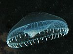 Aequorea victoria (jellyfish) Aequorea victoria (jellyfish)synthetic construct (others) | |||||||||
| Method | ELECTRON MICROSCOPY / single particle reconstruction / cryo EM / Resolution: 3.38 Å | |||||||||
 Authors Authors | Han, S.C. / Li, M.H. | |||||||||
| Funding support | 1items
| |||||||||
 Citation Citation | Journal: To Be Published Title: Cryo-EM structure determination of GPCRs with disulfide-crosslinked fusion protein Authors: Han, S.C. / Li, M.H. | |||||||||
| History |
|
- Structure visualization
| Structure viewer | Molecule: MolmilJmol/JSmol |
|---|
- Downloads & links
-Download
| PDBx/mmCIF format | 9m7g.cif.gz | 166.2 KB | Display | PDBx/mmCIF format |
|---|---|---|---|---|
| PDB format | pdb9m7g.ent.gz | 125 KB | Display | PDB format |
| PDBx/mmJSON format | 9m7g.json.gz | Tree view | PDBx/mmJSON format | |
| Others |  Other downloads Other downloads |
-Validation report
| Arichive directory | https://data.pdbj.org/pub/pdb/validation_reports/m7/9m7gftp://data.pdbj.org/pub/pdb/validation_reports/m7/9m7g | HTTPS FTP |
|---|
-Related structure data
| Related structure data |  63682MC  9m7hC M: map data used to model this data C: citing same article ( |
|---|---|
| Similar structure data |

-Links
 PDBj
PDBj

- Assembly
| Deposited unit | 
|
|---|---|
| 1 |
|
-Components
| #1: Protein | Mass: 53980.281 Da / Num. of mol.: 1 Source method: isolated from a genetically manipulated source Details: Author stated 335-364 is GFP10-11.,Author stated 335-364 is GFP10-11. Source: (gene. exp.) Homo sapiens (human) / Gene: GCGR / Production host: Homo sapiens (human) / References: UniProt: P47871 |
|---|---|
| #2: Protein | Mass: 22388.293 Da / Num. of mol.: 1 Source method: isolated from a genetically manipulated source Details: Author stated 2-3 is GFP1-9. / Source: (gene. exp.) Aequorea victoria (jellyfish) / Gene: gfp / Production host: Homo sapiens (human) / References: UniProt: A0A059PIQ0 |
| #3: Protein | Mass: 31038.664 Da / Num. of mol.: 1 Source method: isolated from a genetically manipulated source Source: (gene. exp.) synthetic construct (others) / Production host: Homo sapiens (human) |
| Has ligand of interest | Y |
| Has protein modification | Y |
-Experimental details
-Experiment
| Experiment | Method: ELECTRON MICROSCOPY |
|---|---|
| EM experiment | Aggregation state: PARTICLE / 3D reconstruction method: single particle reconstruction |
- Sample preparation
| Component | Name: Apo GCGR-GFP10-11:GFP1-9:GFP-clamp E186C complex / Type: COMPLEX / Entity ID: all / Source: RECOMBINANT |
|---|---|
| Source (natural) | Organism: Homo sapiens (human) |
| Source (recombinant) | Organism: Homo sapiens (human) |
| Buffer solution | pH: 7.4 |
| Specimen | Embedding applied: NO / Shadowing applied: NO / Staining applied: NO / Vitrification applied: YES |
| Vitrification | Cryogen name: ETHANE |
- Electron microscopy imaging
| Experimental equipment |  Model: Titan Krios / Image courtesy: FEI Company |
|---|---|
| Microscopy | Model: TFS KRIOS |
| Electron gun | Electron source:  FIELD EMISSION GUN / Accelerating voltage: 300 kV / Illumination mode: FLOOD BEAM FIELD EMISSION GUN / Accelerating voltage: 300 kV / Illumination mode: FLOOD BEAM |
| Electron lens | Mode: BRIGHT FIELD / Nominal defocus max: 2200 nm / Nominal defocus min: 1200 nm |
| Image recording | Electron dose: 60 e/Å2 / Film or detector model: GATAN K2 SUMMIT (4k x 4k) |
- Processing
| EM software |
| ||||||||||||||||||||||||
|---|---|---|---|---|---|---|---|---|---|---|---|---|---|---|---|---|---|---|---|---|---|---|---|---|---|
| CTF correction | Type: NONE | ||||||||||||||||||||||||
| 3D reconstruction | Resolution: 3.38 Å / Resolution method: FSC 0.143 CUT-OFF / Num. of particles: 107457 / Symmetry type: POINT | ||||||||||||||||||||||||
| Refinement | Highest resolution: 3.38 Å Stereochemistry target values: REAL-SPACE (WEIGHTED MAP SUM AT ATOM CENTERS) | ||||||||||||||||||||||||
| Refine LS restraints |
|
