[English] 日本語
 Yorodumi
- PDB-9m5u: Cryo-EM structure of Arabidopsis thaliana MET1 (aa:621-1534) in c... -
+ Open data
- Basic information
| Entry | Database: PDB / ID: 9m5u | |||||||||||||||||||||||||||
|---|---|---|---|---|---|---|---|---|---|---|---|---|---|---|---|---|---|---|---|---|---|---|---|---|---|---|---|---|
| Title | Cryo-EM structure of Arabidopsis thaliana MET1 (aa:621-1534) in complex with hemimethylated DNA analog | |||||||||||||||||||||||||||
 Components Components |
| |||||||||||||||||||||||||||
 Keywords Keywords | STRUCTURAL PROTEIN/DNA / DNA methylation / STRUCTURAL PROTEIN / STRUCTURAL PROTEIN-DNA complex | |||||||||||||||||||||||||||
| Function / homology |  Function and homology information Function and homology informationzygote asymmetric cytokinesis in embryo sac / negative regulation of flower development / DNA-mediated transformation / DNA (cytosine-5-)-methyltransferase / DNA (cytosine-5-)-methyltransferase activity / DNA methylation-dependent constitutive heterochromatin formation / methyltransferase activity / methylation / chromatin binding / DNA binding / nucleus Similarity search - Function | |||||||||||||||||||||||||||
| Biological species |  synthetic construct (others) | |||||||||||||||||||||||||||
| Method | ELECTRON MICROSCOPY / single particle reconstruction / cryo EM / Resolution: 2.74 Å | |||||||||||||||||||||||||||
 Authors Authors | Kikuchi, A. / Arita, K. | |||||||||||||||||||||||||||
| Funding support |  Japan, 1items Japan, 1items
| |||||||||||||||||||||||||||
 Citation Citation |  Journal: Nat Commun / Year: 2025 Journal: Nat Commun / Year: 2025Title: Cryo-EM reveals evolutionarily conserved and distinct structural features of plant CG maintenance methyltransferase MET1. Authors: Amika Kikuchi / Atsuya Nishiyama / Yoshie Chiba / Makoto Nakanishi / Taiko Kim To / Kyohei Arita /  Abstract: DNA methylation is essential for genomic function and transposable element silencing. In plants, DNA methylation occurs in CG, CHG, and CHH contexts (where H = A, T, or C), with the maintenance ...DNA methylation is essential for genomic function and transposable element silencing. In plants, DNA methylation occurs in CG, CHG, and CHH contexts (where H = A, T, or C), with the maintenance of CG methylation mediated by the DNA methyltransferase MET1. The molecular mechanism by which MET1 maintains CG methylation, however, remains unclear. Here, we report cryogenic electron microscopy structures of Arabidopsis thaliana MET1. We find that the methyltransferase domain of MET1 specifically methylates hemimethylated DNA in vitro. The structure of MET1 bound to hemimethylated DNA reveals the activation mechanism of MET1 resembling that of mammalian DNMT1. Curiously, the structure of apo-MET1 shows an autoinhibitory state distinct from that of DNMT1, where the RFTS2 domain and the connecting linker inhibit DNA binding. The autoinhibition of MET1 is relieved upon binding of a potential activator, ubiquitinated histone H3. Taken together, our structural analysis demonstrates both conserved and distinct molecular mechanisms regulating CG maintenance methylation in plant and animal DNA methyltransferases. | |||||||||||||||||||||||||||
| History |
|
- Structure visualization
| Structure viewer | Molecule:  Molmil Molmil Jmol/JSmol Jmol/JSmol |
|---|
- Downloads & links
- Download
| PDBx/mmCIF format |  9m5u.cif.gz 9m5u.cif.gz | 173.3 KB | Display |  PDBx/mmCIF format PDBx/mmCIF format |
|---|---|---|---|---|
| PDB format |  pdb9m5u.ent.gz pdb9m5u.ent.gz | Display |  PDB format PDB format | |
| PDBx/mmJSON format |  9m5u.json.gz 9m5u.json.gz | Tree view |  PDBx/mmJSON format PDBx/mmJSON format | |
| Others |  Other downloads Other downloads |
-Validation report
| Arichive directory |  https://data.pdbj.org/pub/pdb/validation_reports/m5/9m5u https://data.pdbj.org/pub/pdb/validation_reports/m5/9m5u ftp://data.pdbj.org/pub/pdb/validation_reports/m5/9m5u ftp://data.pdbj.org/pub/pdb/validation_reports/m5/9m5u | HTTPS FTP |
|---|
-Related structure data
| Related structure data |  63650MC  9m5xC M: map data used to model this data C: citing same article ( |
|---|---|
| Similar structure data | Similarity search - Function & homology  F&H Search F&H Search |
- Links
Links
- Assembly
| Deposited unit | 
|
|---|---|
| 1 |
|
- Components
| #1: Protein | Mass: 102944.789 Da / Num. of mol.: 1 Source method: isolated from a genetically manipulated source Source: (gene. exp.)  Gene: DMT1, ATHIM, DDM2, DMT01, MET1, MET2, At5g49160, K21P3.3 Production host:  References: UniProt: P34881, DNA (cytosine-5-)-methyltransferase |
|---|---|
| #2: DNA chain | Mass: 3725.469 Da / Num. of mol.: 1 / Source method: obtained synthetically / Source: (synth.) synthetic construct (others) |
| #3: DNA chain | Mass: 3651.431 Da / Num. of mol.: 1 / Source method: obtained synthetically / Source: (synth.) synthetic construct (others) |
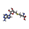
| #4: Chemical | ChemComp-SAH / |
| #5: Chemical | ChemComp-ZN / |
| Has ligand of interest | N |
| Has protein modification | N |
-Experimental details
-Experiment
| Experiment | Method: ELECTRON MICROSCOPY |
|---|---|
| EM experiment | Aggregation state: PARTICLE / 3D reconstruction method: single particle reconstruction |
- Sample preparation
| Component | Name: MET1:DNA / Type: COMPLEX / Entity ID: #1-#3 / Source: MULTIPLE SOURCES |
|---|---|
| Molecular weight | Experimental value: NO |
| Source (natural) | Organism:  |
| Source (recombinant) | Organism:  |
| Buffer solution | pH: 7.5 |
| Specimen | Embedding applied: NO / Shadowing applied: NO / Staining applied: NO / Vitrification applied: YES |
| Vitrification | Cryogen name: ETHANE |
- Electron microscopy imaging
| Experimental equipment |  Model: Titan Krios / Image courtesy: FEI Company |
|---|---|
| Microscopy | Model: TFS KRIOS |
| Electron gun | Electron source:  FIELD EMISSION GUN / Accelerating voltage: 300 kV / Illumination mode: FLOOD BEAM FIELD EMISSION GUN / Accelerating voltage: 300 kV / Illumination mode: FLOOD BEAM |
| Electron lens | Mode: BRIGHT FIELD / Nominal defocus max: 1600 nm / Nominal defocus min: 800 nm |
| Image recording | Electron dose: 60.725 e/Å2 / Film or detector model: GATAN K3 BIOQUANTUM (6k x 4k) |
- Processing
| EM software | Name: PHENIX / Version: 1.20.1_4487 / Category: model refinement | ||||||||||||||||||||||||
|---|---|---|---|---|---|---|---|---|---|---|---|---|---|---|---|---|---|---|---|---|---|---|---|---|---|
| CTF correction | Type: PHASE FLIPPING AND AMPLITUDE CORRECTION | ||||||||||||||||||||||||
| 3D reconstruction | Resolution: 2.74 Å / Resolution method: FSC 0.143 CUT-OFF / Num. of particles: 864086 / Symmetry type: POINT | ||||||||||||||||||||||||
| Refinement | Highest resolution: 2.74 Å Stereochemistry target values: REAL-SPACE (WEIGHTED MAP SUM AT ATOM CENTERS) | ||||||||||||||||||||||||
| Refine LS restraints |
|
 Movie
Movie Controller


 PDBj
PDBj


