 Movie
Movie Controller

[English] 日本語
 Yorodumi
- PDB-9m1e: Crystal structure of the CPS-6 H148A/F122A versus cis-resveratrol... -
+ Open data

- Basic information
| Entry | Database: PDB / ID: 9m1e | ||||||
|---|---|---|---|---|---|---|---|
| Title | Crystal structure of the CPS-6 H148A/F122A versus cis-resveratrol complex | ||||||
 Components Components | Endonuclease G, mitochondrial | ||||||
 Keywords Keywords | DNA BINDING PROTEIN / complex / mitochondrial and endoribonuclease | ||||||
| Function / homology |  Function and homology information Function and homology informationHydrolases; Acting on ester bonds; Endoribonucleases that are active with either ribo- or deoxyribonucleic acids and produce 5'-phosphomonoesters / double-stranded DNA endonuclease activity / single-stranded DNA endonuclease activity / apoptotic DNA fragmentation / DNA catabolic process / RNA catabolic process / RNA endonuclease activity / DNA endonuclease activity / mitochondrial intermembrane space / endonuclease activity ...Hydrolases; Acting on ester bonds; Endoribonucleases that are active with either ribo- or deoxyribonucleic acids and produce 5'-phosphomonoesters / double-stranded DNA endonuclease activity / single-stranded DNA endonuclease activity / apoptotic DNA fragmentation / DNA catabolic process / RNA catabolic process / RNA endonuclease activity / DNA endonuclease activity / mitochondrial intermembrane space / endonuclease activity / sequence-specific DNA binding / mitochondrial inner membrane / protein homodimerization activity / mitochondrion / metal ion binding / nucleus Similarity search - Function | ||||||
| Biological species |  | ||||||
| Method |  X-RAY DIFFRACTION / SYNCHROTRON / MOLECULAR REPLACEMENT / Resolution: 2.99 Å X-RAY DIFFRACTION / SYNCHROTRON / MOLECULAR REPLACEMENT / Resolution: 2.99 Å | ||||||
 Authors Authors | Lin, L.J. / Yuan, H.S. | ||||||
| Funding support |  Taiwan, 1items Taiwan, 1items
| ||||||
 Citation Citation | Journal: to be published Title: Crystal structure of the CPS-6 H148A/F122A versus cis-resveratrol complex Authors: Lin, L.J. / Yuan, H.S. #1: Journal: Nucleic Acids Res. / Year: 2016Title: Crystal structure of endonuclease G in complex with DNA reveals how it nonspecifically degrades DNA as a homodimer Authors: Lin, L.J. / Wu, C.C. / Yang, W.Z. / Yuan, H.S. | ||||||
| History |
|
- Structure visualization
| Structure viewer | Molecule: MolmilJmol/JSmol |
|---|
- Downloads & links
-Download
| PDBx/mmCIF format | 9m1e.cif.gz | 193.7 KB | Display | PDBx/mmCIF format |
|---|---|---|---|---|
| PDB format | pdb9m1e.ent.gz | 156.2 KB | Display | PDB format |
| PDBx/mmJSON format | 9m1e.json.gz | Tree view | PDBx/mmJSON format | |
| Others |  Other downloads Other downloads |
-Validation report
| Arichive directory | https://data.pdbj.org/pub/pdb/validation_reports/m1/9m1eftp://data.pdbj.org/pub/pdb/validation_reports/m1/9m1e | HTTPS FTP |
|---|
-Related structure data
| Related structure data | 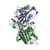 5gkcC 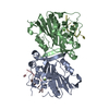 5gkpSC S: Starting model for refinement C: citing same article ( |
|---|---|
| Similar structure data |
-Links
 PDBj
PDBj
- Assembly
| Deposited unit | 
| ||||||||||
|---|---|---|---|---|---|---|---|---|---|---|---|
| 1 |
| ||||||||||
| Unit cell |
|
-Components
| #1: Protein | Mass: 28697.721 Da / Num. of mol.: 2 / Fragment: UNP RESIDUES 63-305 / Mutation: F122A and H148A Source method: isolated from a genetically manipulated source Details: N-terminal 6xhistidine tagged / Source: (gene. exp.)  References: UniProt: Q95NM6, Hydrolases; Acting on ester bonds; Endoribonucleases that are active with either ribo- or deoxyribonucleic acids and produce 5'-phosphomonoesters #2: Chemical | ChemComp-STL /   Mass: 228.243 Da / Num. of mol.: 4 / Source method: obtained synthetically / Formula: C14H12O3 / Feature type: SUBJECT OF INVESTIGATION Mass: 228.243 Da / Num. of mol.: 4 / Source method: obtained synthetically / Formula: C14H12O3 / Feature type: SUBJECT OF INVESTIGATION#3: Chemical |   Mass: 24.305 Da / Num. of mol.: 2 / Source method: obtained synthetically / Formula: Mg / Feature type: SUBJECT OF INVESTIGATION Mass: 24.305 Da / Num. of mol.: 2 / Source method: obtained synthetically / Formula: Mg / Feature type: SUBJECT OF INVESTIGATION#4: Water | ChemComp-HOH / |  Mass: 18.015 Da / Num. of mol.: 4 / Source method: isolated from a natural source / Formula: H2O Mass: 18.015 Da / Num. of mol.: 4 / Source method: isolated from a natural source / Formula: H2OHas ligand of interest | Y | Has protein modification | N | |
|---|
-Experimental details
-Experiment
| Experiment | Method: X-RAY DIFFRACTION / Number of used crystals: 1 |
|---|
- Sample preparation
| Crystal | Density Matthews: 2.35 Å3/Da / Density % sol: 47.6 % / Description: thin plate shape |
|---|---|
| Crystal grow | Temperature: 279 K / Method: vapor diffusion, hanging drop / pH: 7.5 / Details: 100 mM HEPES, 30% (w/v) PEG 1000 |
-Data collection
| Diffraction | Mean temperature: 193 K / Serial crystal experiment: N |
|---|---|
| Diffraction source | Source: SYNCHROTRON / Site: NSRRC / Beamline: TPS 05A / Wavelength: 0.62 Å |
| Detector | Type: RAYONIX MX300-HS / Detector: CCD / Date: Sep 20, 2017 / Details: A Si(111) double-crystal monochromator (DCM) |
| Radiation | Monochromator: LN2 cooled Si(111) double crystal monochromator (DCM) Protocol: SINGLE WAVELENGTH / Monochromatic (M) / Laue (L): M / Scattering type: x-ray |
| Radiation wavelength | Wavelength: 0.62 Å / Relative weight: 1 |
| Reflection | Resolution: 2.99→29.93 Å / Num. obs: 10238 / % possible obs: 99.6 % / Redundancy: 3.6 % / Biso Wilson estimate: 60.77 Å2 / Rpim(I) all: 0.102 / Rrim(I) all: 0.196 / Rsym value: 0.158 / Net I/σ(I): 7.3 |
| Reflection shell | Resolution: 3→3.11 Å / Mean I/σ(I) obs: 2.3 / Num. unique obs: 286093 / Rpim(I) all: 0.313 / Rrim(I) all: 0.573 / Rsym value: 0.456 / Χ2: 0.874 / % possible all: 98 |
- Processing
| Software |
| |||||||||||||||||||||
|---|---|---|---|---|---|---|---|---|---|---|---|---|---|---|---|---|---|---|---|---|---|---|
| Refinement | Method to determine structure: MOLECULAR REPLACEMENT Starting model: 5GKP Resolution: 2.99→29.9 Å / Cross valid method: FREE R-VALUE
| |||||||||||||||||||||
| Refinement step | Cycle: LAST / Resolution: 2.99→29.9 Å
|
