 Movie
Movie Controller


+ Open data

- Basic information
| Entry | Database: PDB / ID: 9lyd | |||||||||
|---|---|---|---|---|---|---|---|---|---|---|
| Title | Cryo-EM structure of GPR3-1IU9 complex | |||||||||
 Components Components | G-protein coupled receptor 3,Aspartate racemase | |||||||||
 Keywords Keywords | STRUCTURAL PROTEIN / GPCR / G protein / cryo-EM / membrane protein | |||||||||
| Function / homology |  Function and homology information Function and homology informationaspartate racemase / aspartate racemase activity / sphingosine-1-phosphate receptor activity / regulation of meiotic nuclear division / G protein-coupled receptor activity / positive regulation of cold-induced thermogenesis / adenylate cyclase-activating G protein-coupled receptor signaling pathway / plasma membrane / cytoplasm Similarity search - Function | |||||||||
| Biological species |  Homo sapiens (human) Homo sapiens (human) | |||||||||
| Method | ELECTRON MICROSCOPY / single particle reconstruction / cryo EM / Resolution: 3.66 Å | |||||||||
 Authors Authors | Hua, T. / Liu, Z.J. / Li, X.T. / Chang, H. | |||||||||
| Funding support |  China, 2items China, 2items
| |||||||||
 Citation Citation | Journal: Cell Rep / Year: 2025 Title: Structural basis of oligomerization-modulated activation and autoinhibition of orphan receptor GPR3. Authors: Hao Chang / Xiaoting Li / Hongqing Tu / Lijie Wu / Yanan Yu / Junlin Liu / Na Chen / Wei L Shen / Tian Hua / Abstract: G protein-coupled receptor 3 (GPR3) is a class A orphan receptor characterized by high constitutive activity in the G signaling pathway. GPR3 has been implicated in Alzheimer's disease and the ...G protein-coupled receptor 3 (GPR3) is a class A orphan receptor characterized by high constitutive activity in the G signaling pathway. GPR3 has been implicated in Alzheimer's disease and the regulation of thermogenesis in human adipocytes, yet the molecular mechanisms underlying its self-activation and potential endogenous modulators remain unclear. In this study, we present cryo-electron microscopy (cryo-EM) structures of GPR3 in different oligomerization states, both in the absence and presence of G protein. Notably, in addition to the monomeric form of GPR3, our findings reveal a functional GPR3 dimer with an extensive dimer interface-a feature rarely observed in class A GPCRs. Moreover, oligomerization appears to be linked to a unique autoinhibition mechanism involving intracellular loops, which may regulate GPR3 signaling. Collectively, these results provide new insights into the oligomerization-modulated activation of orphan GPCRs, advancing our understanding of their signaling properties. | |||||||||
| History |
|
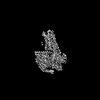
- Structure visualization
| Structure viewer | Molecule: MolmilJmol/JSmol |
|---|
- Downloads & links
-Download
| PDBx/mmCIF format | 9lyd.cif.gz | 137.5 KB | Display | PDBx/mmCIF format |
|---|---|---|---|---|
| PDB format | pdb9lyd.ent.gz | 105.9 KB | Display | PDB format |
| PDBx/mmJSON format | 9lyd.json.gz | Tree view | PDBx/mmJSON format | |
| Others |  Other downloads Other downloads |
-Validation report
| Arichive directory | https://data.pdbj.org/pub/pdb/validation_reports/ly/9lydftp://data.pdbj.org/pub/pdb/validation_reports/ly/9lyd | HTTPS FTP |
|---|
-Related structure data
| Related structure data | 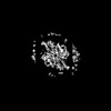 63522MC 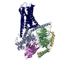 9lybC 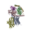 9lycC M: map data used to model this data C: citing same article ( |
|---|---|
| Similar structure data |
-Links
 PDBj
PDBj


- Assembly
| Deposited unit | 
|
|---|---|
| 1 |
|
-Components
| #1: Protein | Mass: 52409.305 Da / Num. of mol.: 2 Source method: isolated from a genetically manipulated source Source: (gene. exp.) Homo sapiens (human) / Gene: GPR3, ACCA, PH0670 / Production host:   Spodoptera frugiperda (fall armyworm) Spodoptera frugiperda (fall armyworm)References: UniProt: P46089, UniProt: O58403, aspartate racemase Has protein modification | Y | |
|---|
-Experimental details
-Experiment
| Experiment | Method: ELECTRON MICROSCOPY |
|---|---|
| EM experiment | Aggregation state: PARTICLE / 3D reconstruction method: single particle reconstruction |
- Sample preparation
| Component | Name: dimer state of GPR3 with PGS / Type: COMPLEX / Entity ID: all / Source: RECOMBINANT |
|---|---|
| Molecular weight | Value: 0.21 MDa / Experimental value: YES |
| Source (natural) | Organism: Homo sapiens (human) |
| Source (recombinant) | Organism: Spodoptera frugiperda (fall armyworm) |
| Buffer solution | pH: 7.5 |
| Specimen | Embedding applied: NO / Shadowing applied: NO / Staining applied: NO / Vitrification applied: YES |
| Specimen support | Grid material: GRAPHENE OXIDE / Grid mesh size: 400 divisions/in. / Grid type: Quantifoil R1.2/1.3 |
| Vitrification | Instrument: FEI VITROBOT MARK IV / Cryogen name: ETHANE |
- Electron microscopy imaging
| Experimental equipment |  Model: Talos Arctica / Image courtesy: FEI Company |
|---|---|
| Microscopy | Model: FEI TECNAI ARCTICA |
| Electron gun | Electron source:  FIELD EMISSION GUN / Accelerating voltage: 300 kV / Illumination mode: FLOOD BEAM FIELD EMISSION GUN / Accelerating voltage: 300 kV / Illumination mode: FLOOD BEAM |
| Electron lens | Mode: BRIGHT FIELD / Nominal defocus max: 2000 nm / Nominal defocus min: 1000 nm / Cs: 2.7 mm / C2 aperture diameter: 50 µm |
| Specimen holder | Cryogen: NITROGEN / Specimen holder model: FEI TITAN KRIOS AUTOGRID HOLDER |
| Image recording | Electron dose: 60 e/Å2 / Film or detector model: FEI FALCON IV (4k x 4k) |
- Processing
| CTF correction | Type: PHASE FLIPPING AND AMPLITUDE CORRECTION |
|---|---|
| Symmetry | Point symmetry: C1 (asymmetric) |
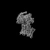
| 3D reconstruction | Resolution: 3.66 Å / Resolution method: FSC 0.143 CUT-OFF / Num. of particles: 205789 / Symmetry type: POINT |
| Atomic model building | B value: 118.6 / Protocol: RIGID BODY FIT |
