 Movie
Movie Controller

[English] 日本語
 Yorodumi
- PDB-9lux: Single-chain Fv antibody of G2 fused with antigen peptide from ch... -
+ Open data

- Basic information
| Entry | Database: PDB / ID: 9lux | ||||||
|---|---|---|---|---|---|---|---|
| Title | Single-chain Fv antibody of G2 fused with antigen peptide from chicken prion protein | ||||||
 Components Components | Major prion protein homolog,Single-chain Fv antibody of G2 fused with antigen peptide from chicken prion protein | ||||||
 Keywords Keywords | IMMUNE SYSTEM / ANTIGEN BINDING / AFFINITY MATURATION / SOMATIC HYPERMUTATION | ||||||
| Function / homology |  Function and homology information Function and homology informationInsertion of tail-anchored proteins into the endoplasmic reticulum membrane / Post-translational modification: synthesis of GPI-anchored proteins / basement membrane / synaptic cleft / side of membrane / protein homooligomerization / copper ion binding / plasma membrane Similarity search - Function | ||||||
| Biological species |   | ||||||
| Method |  X-RAY DIFFRACTION / SYNCHROTRON / MOLECULAR REPLACEMENT / Resolution: 1.85 Å X-RAY DIFFRACTION / SYNCHROTRON / MOLECULAR REPLACEMENT / Resolution: 1.85 Å | ||||||
 Authors Authors | Hanazono, Y. / Yabuno, S. / Hayashi, T. / Numoto, N. / Ito, N. / Oda, M. | ||||||
| Funding support | 1items
| ||||||
 Citation Citation | Journal: Febs Lett. / Year: 2025 Title: Crystal structures reveal how the multispecific antibody G2 achieves binding to different peptides. Authors: Hanazono, Y. / Yabuno, S. / Hayashi, T. / Numoto, N. / Kamatari, Y.O. / Ito, N. / Oda, M. | ||||||
| History |
|
- Structure visualization
| Structure viewer | Molecule: MolmilJmol/JSmol |
|---|
- Downloads & links
-Download
| PDBx/mmCIF format | 9lux.cif.gz | 350.2 KB | Display | PDBx/mmCIF format |
|---|---|---|---|---|
| PDB format | pdb9lux.ent.gz | 258.5 KB | Display | PDB format |
| PDBx/mmJSON format | 9lux.json.gz | Tree view | PDBx/mmJSON format | |
| Others |  Other downloads Other downloads |
-Validation report
| Summary document | 9lux_validation.pdf.gz | 505.8 KB | Display | wwPDB validaton report |
|---|---|---|---|---|
| Full document | 9lux_full_validation.pdf.gz | 523.7 KB | Display | |
| Data in XML | 9lux_validation.xml.gz | 75.3 KB | Display | |
| Data in CIF | 9lux_validation.cif.gz | 99.6 KB | Display | |
| Arichive directory | https://data.pdbj.org/pub/pdb/validation_reports/lu/9luxftp://data.pdbj.org/pub/pdb/validation_reports/lu/9lux | HTTPS FTP |
-Related structure data
| Related structure data |  9luyC 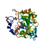 6pgxS S: Starting model for refinement C: citing same article ( |
|---|---|
| Similar structure data |
-Links
 PDBj
PDBj


- Assembly
| Deposited unit | 
| ||||||||||||
|---|---|---|---|---|---|---|---|---|---|---|---|---|---|
| 1 | 
| ||||||||||||
| 2 | 
| ||||||||||||
| 3 | 
| ||||||||||||
| 4 | 
| ||||||||||||
| 5 | 
| ||||||||||||
| 6 | 
| ||||||||||||
| Unit cell |
|
-Components
| #1: Antibody | Mass: 28806.979 Da / Num. of mol.: 6 Source method: isolated from a genetically manipulated source Details: The fusion protein composed of an expression tag (residues 1-2), a fragment from chicken prion protein (residues 3-20), a linker (residues 21-28), the light chain (residues 29-138), a second ...Details: The fusion protein composed of an expression tag (residues 1-2), a fragment from chicken prion protein (residues 3-20), a linker (residues 21-28), the light chain (residues 29-138), a second linker (residues 139-153), and the heavy chain (residues 154-271). Source: (gene. exp.) Gene: PRNP, PRN-P / Production host:  #2: Chemical | ChemComp-SO4 /   Mass: 96.063 Da / Num. of mol.: 15 / Source method: obtained synthetically / Formula: SO4 Mass: 96.063 Da / Num. of mol.: 15 / Source method: obtained synthetically / Formula: SO4#3: Chemical | ChemComp-GOL /   Mass: 92.094 Da / Num. of mol.: 4 / Source method: obtained synthetically / Formula: C3H8O3 Mass: 92.094 Da / Num. of mol.: 4 / Source method: obtained synthetically / Formula: C3H8O3#4: Water | ChemComp-HOH / |  Mass: 18.015 Da / Num. of mol.: 982 / Source method: isolated from a natural source / Formula: H2O Mass: 18.015 Da / Num. of mol.: 982 / Source method: isolated from a natural source / Formula: H2OHas ligand of interest | N | Has protein modification | Y | |
|---|
-Experimental details
-Experiment
| Experiment | Method: X-RAY DIFFRACTION / Number of used crystals: 1 |
|---|
- Sample preparation
| Crystal | Density Matthews: 3.29 Å3/Da / Density % sol: 62.67 % |
|---|---|
| Crystal grow | Temperature: 293 K / Method: vapor diffusion, sitting drop Details: 0.5 M ammonium sulphate, 0.1 M sodium citrate tribasic dihydrate pH 5.6, 1.0 M lithium sulfate monohydrate |
-Data collection
| Diffraction | Mean temperature: 100 K / Serial crystal experiment: N |
|---|---|
| Diffraction source | Source: SYNCHROTRON / Site: SLS  / Beamline: X06SA / Wavelength: 1 Å / Beamline: X06SA / Wavelength: 1 Å |
| Detector | Type: DECTRIS EIGER X 16M / Detector: PIXEL / Date: Aug 18, 2021 |
| Radiation | Protocol: SINGLE WAVELENGTH / Monochromatic (M) / Laue (L): M / Scattering type: x-ray |
| Radiation wavelength | Wavelength: 1 Å / Relative weight: 1 |
| Reflection | Resolution: 1.85→50 Å / Num. obs: 183582 / % possible obs: 97.3 % / Redundancy: 3.2 % / Biso Wilson estimate: 33.2 Å2 / Rmerge(I) obs: 0.094 / Net I/σ(I): 8.5 |
| Reflection shell | Resolution: 1.85→1.96 Å / Redundancy: 3.2 % / Rmerge(I) obs: 0.786 / Mean I/σ(I) obs: 1.8 / Num. unique obs: 29434 / CC1/2: 0.604 |
- Processing
| Software |
| |||||||||||||||||||||||||||||||||||||||||||||||||||||||||||||||||||||||||||||||||||||||||||||||||||||||||||||||||||||||||||||||||||||||||||||||||||||||||||||||||||||||||||||||||||||||||||||||||||||||||||||||||||||||||
|---|---|---|---|---|---|---|---|---|---|---|---|---|---|---|---|---|---|---|---|---|---|---|---|---|---|---|---|---|---|---|---|---|---|---|---|---|---|---|---|---|---|---|---|---|---|---|---|---|---|---|---|---|---|---|---|---|---|---|---|---|---|---|---|---|---|---|---|---|---|---|---|---|---|---|---|---|---|---|---|---|---|---|---|---|---|---|---|---|---|---|---|---|---|---|---|---|---|---|---|---|---|---|---|---|---|---|---|---|---|---|---|---|---|---|---|---|---|---|---|---|---|---|---|---|---|---|---|---|---|---|---|---|---|---|---|---|---|---|---|---|---|---|---|---|---|---|---|---|---|---|---|---|---|---|---|---|---|---|---|---|---|---|---|---|---|---|---|---|---|---|---|---|---|---|---|---|---|---|---|---|---|---|---|---|---|---|---|---|---|---|---|---|---|---|---|---|---|---|---|---|---|---|---|---|---|---|---|---|---|---|---|---|---|---|---|---|---|---|
| Refinement | Method to determine structure: MOLECULAR REPLACEMENT Starting model: 6PGX Resolution: 1.85→49.32 Å / SU ML: 0.2466 / Cross valid method: FREE R-VALUE / σ(F): 1.96 / Phase error: 24.7367 Stereochemistry target values: GeoStd + Monomer Library + CDL v1.2
| |||||||||||||||||||||||||||||||||||||||||||||||||||||||||||||||||||||||||||||||||||||||||||||||||||||||||||||||||||||||||||||||||||||||||||||||||||||||||||||||||||||||||||||||||||||||||||||||||||||||||||||||||||||||||
| Solvent computation | Shrinkage radii: 0.9 Å / VDW probe radii: 1.1 Å / Solvent model: FLAT BULK SOLVENT MODEL | |||||||||||||||||||||||||||||||||||||||||||||||||||||||||||||||||||||||||||||||||||||||||||||||||||||||||||||||||||||||||||||||||||||||||||||||||||||||||||||||||||||||||||||||||||||||||||||||||||||||||||||||||||||||||
| Displacement parameters | Biso mean: 39.16 Å2 | |||||||||||||||||||||||||||||||||||||||||||||||||||||||||||||||||||||||||||||||||||||||||||||||||||||||||||||||||||||||||||||||||||||||||||||||||||||||||||||||||||||||||||||||||||||||||||||||||||||||||||||||||||||||||
| Refinement step | Cycle: LAST / Resolution: 1.85→49.32 Å
| |||||||||||||||||||||||||||||||||||||||||||||||||||||||||||||||||||||||||||||||||||||||||||||||||||||||||||||||||||||||||||||||||||||||||||||||||||||||||||||||||||||||||||||||||||||||||||||||||||||||||||||||||||||||||
| Refine LS restraints |
| |||||||||||||||||||||||||||||||||||||||||||||||||||||||||||||||||||||||||||||||||||||||||||||||||||||||||||||||||||||||||||||||||||||||||||||||||||||||||||||||||||||||||||||||||||||||||||||||||||||||||||||||||||||||||
| LS refinement shell |
|
