 Movie
Movie Controller

[English] 日本語
 Yorodumi
- PDB-9lns: Crystal structure of de novo designed amantadine induced homotrim... -
+ Open data

- Basic information
| Entry | Database: PDB / ID: 9lns | ||||||
|---|---|---|---|---|---|---|---|
| Title | Crystal structure of de novo designed amantadine induced homotrimer dAIT17 | ||||||
 Components Components | dAIT17 | ||||||
 Keywords Keywords | DE NOVO PROTEIN / Drug induced homotrimer | ||||||
| Function / homology | (3S,5S,7S)-tricyclo[3.3.1.1~3,7~]decan-1-amine Function and homology information Function and homology information | ||||||
| Biological species |  | ||||||
| Method |  X-RAY DIFFRACTION / MOLECULAR REPLACEMENT / Resolution: 1.5 Å X-RAY DIFFRACTION / MOLECULAR REPLACEMENT / Resolution: 1.5 Å | ||||||
 Authors Authors | Qihan, J. / Longxing, C. | ||||||
| Funding support |  China, 1items China, 1items
| ||||||
 Citation Citation | Journal: Science / Year: 2026 Title: De novo design of small molecule-regulated protein oligomers. Authors: Jin, Q. / Wang, Y. / Chen, D. / Liao, J. / Cui, Z. / Fan, Y. / Zeng, A. / Xie, M. / Cao, L. | ||||||
| History |
|
- Structure visualization
| Structure viewer | Molecule: MolmilJmol/JSmol |
|---|
- Downloads & links
-Download
| PDBx/mmCIF format | 9lns.cif.gz | 28.5 KB | Display | PDBx/mmCIF format |
|---|---|---|---|---|
| PDB format | pdb9lns.ent.gz | 17.8 KB | Display | PDB format |
| PDBx/mmJSON format | 9lns.json.gz | Tree view | PDBx/mmJSON format | |
| Others |  Other downloads Other downloads |
-Validation report
| Arichive directory | https://data.pdbj.org/pub/pdb/validation_reports/ln/9lnsftp://data.pdbj.org/pub/pdb/validation_reports/ln/9lns | HTTPS FTP |
|---|
-Related structure data


-Links
 PDBj
PDBj
- Assembly
| Deposited unit | 
| |||||||||||||||||||||
|---|---|---|---|---|---|---|---|---|---|---|---|---|---|---|---|---|---|---|---|---|---|---|
| 1 |
| |||||||||||||||||||||
| Unit cell |
| |||||||||||||||||||||
| Components on special symmetry positions |
|
-Components
| #1: Protein | Mass: 7463.866 Da / Num. of mol.: 1 Source method: isolated from a genetically manipulated source Source: (gene. exp.) | ||||||
|---|---|---|---|---|---|---|---|
| #2: Chemical | 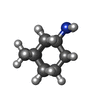  Mass: 151.249 Da / Num. of mol.: 2 / Source method: obtained synthetically / Formula: C10H17N / Feature type: SUBJECT OF INVESTIGATION Mass: 151.249 Da / Num. of mol.: 2 / Source method: obtained synthetically / Formula: C10H17N / Feature type: SUBJECT OF INVESTIGATION#3: Water | ChemComp-HOH / |  Mass: 18.015 Da / Num. of mol.: 111 / Source method: isolated from a natural source / Formula: H2O Mass: 18.015 Da / Num. of mol.: 111 / Source method: isolated from a natural source / Formula: H2OHas ligand of interest | Y | Has protein modification | N | |
-Experimental details
-Experiment
| Experiment | Method: X-RAY DIFFRACTION / Number of used crystals: 1 |
|---|
- Sample preparation
| Crystal | Density Matthews: 2.8 Å3/Da / Density % sol: 56 % |
|---|---|
| Crystal grow | Temperature: 293.15 K / Method: vapor diffusion, sitting drop / pH: 5.5 Details: 0.1 M Sodium citrate tribasic dihydrate pH 5.5, 22% w/v Polyethylene glycol 1,000 |
-Data collection
| Diffraction | Mean temperature: 181 K / Ambient temp details: 100 / Serial crystal experiment: N |
|---|---|
| Diffraction source | Source: ROTATING ANODE / Type: Cu FINE FOCUS / Wavelength: 1 Å |
| Detector | Type: RIGAKU HyPix-6000HE / Detector: PIXEL / Date: Mar 23, 2024 |
| Radiation | Protocol: SINGLE WAVELENGTH / Monochromatic (M) / Laue (L): M / Scattering type: x-ray |
| Radiation wavelength | Wavelength: 1 Å / Relative weight: 1 |
| Reflection | Resolution: 1.5→27.99 Å / Num. obs: 14121 / % possible obs: 99.67 % / Redundancy: 3.7 % / CC1/2: 1 / Net I/σ(I): 22.56 |
| Reflection shell | Resolution: 1.5→1.55 Å / Num. unique obs: 1374 / CC1/2: 0.665 |
- Processing
| Software |
| |||||||||||||||||||||||||||||||||||||||||||||||||||||||||||||||||||||||||||||
|---|---|---|---|---|---|---|---|---|---|---|---|---|---|---|---|---|---|---|---|---|---|---|---|---|---|---|---|---|---|---|---|---|---|---|---|---|---|---|---|---|---|---|---|---|---|---|---|---|---|---|---|---|---|---|---|---|---|---|---|---|---|---|---|---|---|---|---|---|---|---|---|---|---|---|---|---|---|---|
| Refinement | Method to determine structure: MOLECULAR REPLACEMENT / Resolution: 1.5→27.99 Å / SU ML: 0.17 / Cross valid method: FREE R-VALUE / σ(F): 1.34 / Phase error: 24.16 / Stereochemistry target values: ML
| |||||||||||||||||||||||||||||||||||||||||||||||||||||||||||||||||||||||||||||
| Solvent computation | Shrinkage radii: 0.9 Å / VDW probe radii: 1.1 Å / Solvent model: FLAT BULK SOLVENT MODEL | |||||||||||||||||||||||||||||||||||||||||||||||||||||||||||||||||||||||||||||
| Refinement step | Cycle: LAST / Resolution: 1.5→27.99 Å
| |||||||||||||||||||||||||||||||||||||||||||||||||||||||||||||||||||||||||||||
| Refine LS restraints |
| |||||||||||||||||||||||||||||||||||||||||||||||||||||||||||||||||||||||||||||
| LS refinement shell |
|
