 Movie
Movie Controller

[English] 日本語
 Yorodumi
- PDB-9ll3: X-ray structure of Enterobacter cloaca transaldolase in complex w... -
+ Open data

- Basic information
| Entry | Database: PDB / ID: 9ll3 | ||||||
|---|---|---|---|---|---|---|---|
| Title | X-ray structure of Enterobacter cloaca transaldolase in complex with D-fructose-6-phosphate. | ||||||
 Components Components | Fructose-6-phosphate aldolase | ||||||
 Keywords Keywords | TRANSFERASE / transaldolase | ||||||
| Function / homology |  Function and homology information Function and homology informationketone catabolic process / fructose 6-phosphate aldolase activity / Lyases; Carbon-carbon lyases; Aldehyde-lyases / fructose metabolic process / cytoplasm Similarity search - Function | ||||||
| Biological species |  Enterobacter cloacae (bacteria) Enterobacter cloacae (bacteria) | ||||||
| Method |  X-RAY DIFFRACTION / MOLECULAR REPLACEMENT / Resolution: 1.92 Å X-RAY DIFFRACTION / MOLECULAR REPLACEMENT / Resolution: 1.92 Å | ||||||
 Authors Authors | Kamitori, S. | ||||||
| Funding support | 1items
| ||||||
 Citation Citation | Journal: J.Agric.Food Chem. / Year: 2025 Title: Synthetic Study of 8- and 9-Carbon Sugars by Transaldolase. Authors: Yoshihara, A. / Miyoshi, E. / Tomino, S. / Hanaki, Y. / Mochizuki, S. / Yoshida, H. / Izumori, K. / Kamitori, S. | ||||||
| History |
|
- Structure visualization
| Structure viewer | Molecule: MolmilJmol/JSmol |
|---|
- Downloads & links
-Download
| PDBx/mmCIF format | 9ll3.cif.gz | 399.5 KB | Display | PDBx/mmCIF format |
|---|---|---|---|---|
| PDB format | pdb9ll3.ent.gz | 326.5 KB | Display | PDB format |
| PDBx/mmJSON format | 9ll3.json.gz | Tree view | PDBx/mmJSON format | |
| Others |  Other downloads Other downloads |
-Validation report
| Arichive directory | https://data.pdbj.org/pub/pdb/validation_reports/ll/9ll3ftp://data.pdbj.org/pub/pdb/validation_reports/ll/9ll3 | HTTPS FTP |
|---|
-Related structure data
| Related structure data |  9lkpC  9ui2C  3s1vS S: Starting model for refinement C: citing same article ( |
|---|---|
| Similar structure data |
-Links
 PDBj
PDBj- Assembly
| Deposited unit | 
| ||||||||
|---|---|---|---|---|---|---|---|---|---|
| 1 | 
| ||||||||
| Unit cell |
| ||||||||
| Components on special symmetry positions |
|
-Components
| #1: Protein | Mass: 25331.182 Da / Num. of mol.: 5 Source method: isolated from a genetically manipulated source Source: (gene. exp.) Enterobacter cloacae (bacteria) / Gene: fsa, DP202_18755 / Production host: References: UniProt: A0A330G8J7, Lyases; Carbon-carbon lyases; Aldehyde-lyases #2: Chemical | ChemComp-F6R / 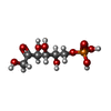  Mass: 260.136 Da / Num. of mol.: 5 / Source method: obtained synthetically / Formula: C6H13O9P / Feature type: SUBJECT OF INVESTIGATION Mass: 260.136 Da / Num. of mol.: 5 / Source method: obtained synthetically / Formula: C6H13O9P / Feature type: SUBJECT OF INVESTIGATION#3: Water | ChemComp-HOH / |  Mass: 18.015 Da / Num. of mol.: 661 / Source method: isolated from a natural source / Formula: H2O Mass: 18.015 Da / Num. of mol.: 661 / Source method: isolated from a natural source / Formula: H2OHas ligand of interest | Y | Has protein modification | Y | |
|---|
-Experimental details
-Experiment
| Experiment | Method: X-RAY DIFFRACTION / Number of used crystals: 1 |
|---|
- Sample preparation
| Crystal | Density Matthews: 2.34 Å3/Da / Density % sol: 47.4 % |
|---|---|
| Crystal grow | Temperature: 298 K / Method: vapor diffusion, sitting drop Details: 100 mM cacodylate buffer, pH 6.5, 10% (w/v) PEG3000, 10% (w/v) PEG 8000, 200 mM of MgCl2, 20 mM dihydroxtacetone, 20 mM, 20 mM D,L-glyceraldehyde-3-phosphate |
-Data collection
| Diffraction | Mean temperature: 100 K / Serial crystal experiment: N |
|---|---|
| Diffraction source | Source: ROTATING ANODE / Type: RIGAKU MICROMAX-007 HF / Wavelength: 1.5418 Å |
| Detector | Type: RIGAKU RAXIS VII / Detector: IMAGE PLATE / Date: Dec 12, 2019 |
| Radiation | Protocol: SINGLE WAVELENGTH / Monochromatic (M) / Laue (L): M / Scattering type: x-ray |
| Radiation wavelength | Wavelength: 1.5418 Å / Relative weight: 1 |
| Reflection | Resolution: 1.92→19.6 Å / Num. obs: 90439 / % possible obs: 99.8 % / Redundancy: 7.2 % / CC1/2: 0.998 / Rmerge(I) obs: 0.117 / Net I/σ(I): 14.7 |
| Reflection shell | Resolution: 1.92→1.97 Å / Redundancy: 7.2 % / Rmerge(I) obs: 0.912 / Mean I/σ(I) obs: 2.3 / Num. unique obs: 6596 / CC1/2: 0.771 |
- Processing
| Software |
| ||||||||||||||||||||||||||||||||||||||||||||||||||||||||||||||||||||||||||||||||||||||||||||||||||||||||||||||||||||||||||||||||||||||||||||||||||||||||||||||||||||||||||||||||||||||
|---|---|---|---|---|---|---|---|---|---|---|---|---|---|---|---|---|---|---|---|---|---|---|---|---|---|---|---|---|---|---|---|---|---|---|---|---|---|---|---|---|---|---|---|---|---|---|---|---|---|---|---|---|---|---|---|---|---|---|---|---|---|---|---|---|---|---|---|---|---|---|---|---|---|---|---|---|---|---|---|---|---|---|---|---|---|---|---|---|---|---|---|---|---|---|---|---|---|---|---|---|---|---|---|---|---|---|---|---|---|---|---|---|---|---|---|---|---|---|---|---|---|---|---|---|---|---|---|---|---|---|---|---|---|---|---|---|---|---|---|---|---|---|---|---|---|---|---|---|---|---|---|---|---|---|---|---|---|---|---|---|---|---|---|---|---|---|---|---|---|---|---|---|---|---|---|---|---|---|---|---|---|---|---|
| Refinement | Method to determine structure: MOLECULAR REPLACEMENT Starting model: 3S1V Resolution: 1.92→19.6 Å / Cor.coef. Fo:Fc: 0.961 / Cor.coef. Fo:Fc free: 0.944 / SU B: 5.816 / SU ML: 0.087 / Cross valid method: THROUGHOUT / ESU R: 0.135 / ESU R Free: 0.126 / Stereochemistry target values: MAXIMUM LIKELIHOOD / Details: HYDROGENS HAVE BEEN ADDED IN THE RIDING POSITIONS
| ||||||||||||||||||||||||||||||||||||||||||||||||||||||||||||||||||||||||||||||||||||||||||||||||||||||||||||||||||||||||||||||||||||||||||||||||||||||||||||||||||||||||||||||||||||||
| Solvent computation | Ion probe radii: 0.8 Å / Shrinkage radii: 0.8 Å / VDW probe radii: 1.2 Å / Solvent model: MASK | ||||||||||||||||||||||||||||||||||||||||||||||||||||||||||||||||||||||||||||||||||||||||||||||||||||||||||||||||||||||||||||||||||||||||||||||||||||||||||||||||||||||||||||||||||||||
| Displacement parameters | Biso mean: 20.293 Å2
| ||||||||||||||||||||||||||||||||||||||||||||||||||||||||||||||||||||||||||||||||||||||||||||||||||||||||||||||||||||||||||||||||||||||||||||||||||||||||||||||||||||||||||||||||||||||
| Refinement step | Cycle: 1 / Resolution: 1.92→19.6 Å
| ||||||||||||||||||||||||||||||||||||||||||||||||||||||||||||||||||||||||||||||||||||||||||||||||||||||||||||||||||||||||||||||||||||||||||||||||||||||||||||||||||||||||||||||||||||||
| Refine LS restraints |
|