 Movie
Movie Controller

[English] 日本語
 Yorodumi
- PDB-9lb7: Crystal structure of trehalose-6-phosphate phosphorylase from Wei... -
+ Open data

- Basic information
| Entry | Database: PDB / ID: 9lb7 | ||||||
|---|---|---|---|---|---|---|---|
| Title | Crystal structure of trehalose-6-phosphate phosphorylase from Weissella ceti in complex with beta-Glc1P | ||||||
 Components Components | Trehalose-6-phosphate hydrolase | ||||||
 Keywords Keywords | HYDROLASE / trehalose-6-phosphate phosphorylase | ||||||
| Function / homology |  Function and homology information Function and homology informationglycosyltransferase activity / hydrolase activity, hydrolyzing O-glycosyl compounds / carbohydrate binding / carbohydrate metabolic process Similarity search - Function | ||||||
| Biological species |  Weissella ceti (bacteria) Weissella ceti (bacteria) | ||||||
| Method |  X-RAY DIFFRACTION / SYNCHROTRON / MOLECULAR REPLACEMENT / Resolution: 2.35 Å X-RAY DIFFRACTION / SYNCHROTRON / MOLECULAR REPLACEMENT / Resolution: 2.35 Å | ||||||
 Authors Authors | Feng, Y. / Xue, S. | ||||||
| Funding support |  China, 1items China, 1items
| ||||||
 Citation Citation | Journal: J.Agric.Food Chem. / Year: 2025 Title: Structural Insights into Trehalose-6-Phosphate Phosphorylase and Its Role in Trehalose 6-Phosphate Biosynthesis via a Multienzyme Cascade. Authors: Feng, Y. / Wang, N. / Cao, Q. / Li, S. / Xu, Y. / Che, X. / Zhao, J. / Yang, C. / Xue, S. | ||||||
| History |
|
- Structure visualization
| Structure viewer | Molecule: MolmilJmol/JSmol |
|---|
- Downloads & links
-Download
| PDBx/mmCIF format | 9lb7.cif.gz | 211.5 KB | Display | PDBx/mmCIF format |
|---|---|---|---|---|
| PDB format | pdb9lb7.ent.gz | 131.6 KB | Display | PDB format |
| PDBx/mmJSON format | 9lb7.json.gz | Tree view | PDBx/mmJSON format | |
| Others |  Other downloads Other downloads |
-Validation report
| Arichive directory | https://data.pdbj.org/pub/pdb/validation_reports/lb/9lb7ftp://data.pdbj.org/pub/pdb/validation_reports/lb/9lb7 | HTTPS FTP |
|---|
-Related structure data

-Links
 PDBj
PDBj


- Assembly
| Deposited unit | 
| ||||||||||||
|---|---|---|---|---|---|---|---|---|---|---|---|---|---|
| 1 |
| ||||||||||||
| Unit cell |
|
-Components
| #1: Protein | Mass: 86645.453 Da / Num. of mol.: 1 Source method: isolated from a genetically manipulated source Source: (gene. exp.) Weissella ceti (bacteria) / Gene: WS74_1295 / Production host: |
|---|---|
| #2: Sugar | ChemComp-XGP / 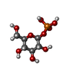  Type: D-saccharide / Mass: 260.136 Da / Num. of mol.: 1 / Source method: obtained synthetically / Formula: C6H13O9P / Feature type: SUBJECT OF INVESTIGATION Type: D-saccharide / Mass: 260.136 Da / Num. of mol.: 1 / Source method: obtained synthetically / Formula: C6H13O9P / Feature type: SUBJECT OF INVESTIGATION |
| #3: Chemical | ChemComp-MG /   Mass: 24.305 Da / Num. of mol.: 1 / Source method: obtained synthetically / Formula: Mg Mass: 24.305 Da / Num. of mol.: 1 / Source method: obtained synthetically / Formula: Mg |
| #4: Water | ChemComp-HOH /  Mass: 18.015 Da / Num. of mol.: 331 / Source method: isolated from a natural source / Formula: H2O Mass: 18.015 Da / Num. of mol.: 331 / Source method: isolated from a natural source / Formula: H2O |
| Has ligand of interest | Y |
| Has protein modification | N |
-Experimental details
-Experiment
| Experiment | Method: X-RAY DIFFRACTION / Number of used crystals: 1 |
|---|
- Sample preparation
| Crystal | Density Matthews: 2.42 Å3/Da / Density % sol: 49.07 % |
|---|---|
| Crystal grow | Temperature: 300 K / Method: vapor diffusion, hanging drop / pH: 6.5 Details: 0.2 M calcium acetate hydrate, 0.1 M sodium cacodylate trihydrate (pH 6.5), and 18% (w/v) polyethylene glycol 8,000 |
-Data collection
| Diffraction | Mean temperature: 100 K / Serial crystal experiment: N |
|---|---|
| Diffraction source | Source: SYNCHROTRON / Site: SSRF / Beamline: BL18U1 / Wavelength: 0.9792 Å |
| Detector | Type: DECTRIS PILATUS 6M / Detector: PIXEL / Date: Oct 9, 2024 |
| Radiation | Protocol: SINGLE WAVELENGTH / Monochromatic (M) / Laue (L): M / Scattering type: x-ray |
| Radiation wavelength | Wavelength: 0.9792 Å / Relative weight: 1 |
| Reflection | Resolution: 2.35→48.21 Å / Num. obs: 34466 / % possible obs: 99.97 % / Redundancy: 5.9 % / Biso Wilson estimate: 30.53 Å2 / CC1/2: 0.973 / Rmerge(I) obs: 0.195 / Net I/σ(I): 10.6 |
| Reflection shell | Resolution: 2.35→2.43 Å / Redundancy: 6.1 % / Rmerge(I) obs: 0.609 / Mean I/σ(I) obs: 3.3 / Num. unique obs: 1710 / CC1/2: 0.816 / % possible all: 88.01 |
- Processing
| Software |
| |||||||||||||||||||||||||||||||||||||||||||||||||||||||||||||||||||||||||||||||||||||||||||||||||||||||||
|---|---|---|---|---|---|---|---|---|---|---|---|---|---|---|---|---|---|---|---|---|---|---|---|---|---|---|---|---|---|---|---|---|---|---|---|---|---|---|---|---|---|---|---|---|---|---|---|---|---|---|---|---|---|---|---|---|---|---|---|---|---|---|---|---|---|---|---|---|---|---|---|---|---|---|---|---|---|---|---|---|---|---|---|---|---|---|---|---|---|---|---|---|---|---|---|---|---|---|---|---|---|---|---|---|---|---|
| Refinement | Method to determine structure: MOLECULAR REPLACEMENT / Resolution: 2.35→48.21 Å / SU ML: 0.3122 / Cross valid method: FREE R-VALUE / σ(F): 1.34 / Phase error: 30.0107 Stereochemistry target values: GeoStd + Monomer Library + CDL v1.2
| |||||||||||||||||||||||||||||||||||||||||||||||||||||||||||||||||||||||||||||||||||||||||||||||||||||||||
| Solvent computation | Shrinkage radii: 0.9 Å / VDW probe radii: 1.1 Å / Solvent model: FLAT BULK SOLVENT MODEL | |||||||||||||||||||||||||||||||||||||||||||||||||||||||||||||||||||||||||||||||||||||||||||||||||||||||||
| Displacement parameters | Biso mean: 33.47 Å2 | |||||||||||||||||||||||||||||||||||||||||||||||||||||||||||||||||||||||||||||||||||||||||||||||||||||||||
| Refinement step | Cycle: LAST / Resolution: 2.35→48.21 Å
| |||||||||||||||||||||||||||||||||||||||||||||||||||||||||||||||||||||||||||||||||||||||||||||||||||||||||
| Refine LS restraints |
| |||||||||||||||||||||||||||||||||||||||||||||||||||||||||||||||||||||||||||||||||||||||||||||||||||||||||
| LS refinement shell |
|
