 Movie
Movie Controller

[English] 日本語
 Yorodumi
- PDB-9l8k: Rhodothermus marines cellobiose 2-epimerase RmCE in complex with ... -
+ Open data

- Basic information
| Entry | Database: PDB / ID: 9l8k | ||||||
|---|---|---|---|---|---|---|---|
| Title | Rhodothermus marines cellobiose 2-epimerase RmCE in complex with mannobiitol | ||||||
 Components Components | Cellobiose 2-epimerase | ||||||
 Keywords Keywords | ISOMERASE / cellobiose 2-epimerase / mannobiitol / epimerization | ||||||
| Function / homology |  Function and homology information Function and homology informationcellobiose epimerase / cellobiose epimerase activity / carbohydrate metabolic process Similarity search - Function | ||||||
| Biological species |   Rhodothermus marinus JCM 9785 (bacteria) Rhodothermus marinus JCM 9785 (bacteria) | ||||||
| Method |  X-RAY DIFFRACTION / SYNCHROTRON / MOLECULAR REPLACEMENT / Resolution: 1.7 Å X-RAY DIFFRACTION / SYNCHROTRON / MOLECULAR REPLACEMENT / Resolution: 1.7 Å | ||||||
 Authors Authors | Saburi, W. / Muto, H. / Jaito, N. / Kato, K. / Yu, J. / Yao, M. / Mori, H. | ||||||
| Funding support | 1items
| ||||||
 Citation Citation | Journal: Biosci.Biotechnol.Biochem. / Year: 2025 Title: Biochemical and structural analysis of the mechanism for the catalysis and specificity of cellobiose 2-epimerase from Rhodothermus marinus. Authors: Saburi, W. / Muto-Fukiya, H. / Jaito, N. / Kato, K. / Yu, J. / Yao, M. / Mori, H. | ||||||
| History |
|
- Structure visualization
| Structure viewer | Molecule: MolmilJmol/JSmol |
|---|
- Downloads & links
-Download
| PDBx/mmCIF format | 9l8k.cif.gz | 101 KB | Display | PDBx/mmCIF format |
|---|---|---|---|---|
| PDB format | pdb9l8k.ent.gz | 74.7 KB | Display | PDB format |
| PDBx/mmJSON format | 9l8k.json.gz | Tree view | PDBx/mmJSON format | |
| Others |  Other downloads Other downloads |
-Validation report
| Arichive directory | https://data.pdbj.org/pub/pdb/validation_reports/l8/9l8kftp://data.pdbj.org/pub/pdb/validation_reports/l8/9l8k | HTTPS FTP |
|---|
-Related structure data
| Related structure data |  9l8iC 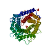 3wkfS S: Starting model for refinement C: citing same article ( |
|---|---|
| Similar structure data |
-Links
 PDBj
PDBj
- Assembly
| Deposited unit | 
| ||||||||
|---|---|---|---|---|---|---|---|---|---|
| 1 |
| ||||||||
| Unit cell |
|
-Components
-Protein / Sugars , 2 types, 2 molecules A
| #1: Protein | Mass: 47386.258 Da / Num. of mol.: 1 Source method: isolated from a genetically manipulated source Details: H36P conflict: error in database Source: (gene. exp.) Rhodothermus marinus JCM 9785 (bacteria)Gene: ce / Production host: |
|---|---|
| #3: Sugar | ChemComp-BMA /  Type: D-saccharide, beta linking / Mass: 180.156 Da / Num. of mol.: 1 / Source method: obtained synthetically / Formula: C6H12O6 Type: D-saccharide, beta linking / Mass: 180.156 Da / Num. of mol.: 1 / Source method: obtained synthetically / Formula: C6H12O6 |
-Non-polymers , 4 types, 246 molecules 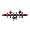


| #2: Chemical | ChemComp-MTL /  Mass: 182.172 Da / Num. of mol.: 1 / Source method: obtained synthetically / Formula: C6H14O6 / Feature type: SUBJECT OF INVESTIGATION / Comment: medication*YM Mass: 182.172 Da / Num. of mol.: 1 / Source method: obtained synthetically / Formula: C6H14O6 / Feature type: SUBJECT OF INVESTIGATION / Comment: medication*YM |
|---|---|
| #4: Chemical | ChemComp-CL /  Mass: 35.453 Da / Num. of mol.: 1 / Source method: obtained synthetically / Formula: Cl Mass: 35.453 Da / Num. of mol.: 1 / Source method: obtained synthetically / Formula: Cl |
| #5: Chemical | ChemComp-PO4 /  Mass: 94.971 Da / Num. of mol.: 1 / Source method: obtained synthetically / Formula: PO4 Mass: 94.971 Da / Num. of mol.: 1 / Source method: obtained synthetically / Formula: PO4 |
| #6: Water | ChemComp-HOH / Mass: 18.015 Da / Num. of mol.: 243 / Source method: isolated from a natural source / Formula: H2O |
-Details
| Has ligand of interest | Y |
|---|---|
| Has protein modification | N |
-Experimental details
-Experiment
| Experiment | Method: X-RAY DIFFRACTION / Number of used crystals: 1 |
|---|
- Sample preparation
| Crystal grow | Temperature: 293 K / Method: vapor diffusion, hanging drop Details: 10 mM mannobiitol, 48 mM NaCl, 100 mM sodium acetate buffer (pH 4.5), 1.2 M ammonium sulfate |
|---|
-Data collection
| Diffraction | Mean temperature: 100 K / Serial crystal experiment: N |
|---|---|
| Diffraction source | Source: SYNCHROTRON / Site: Photon Factory  / Beamline: BL-1A / Wavelength: 1.1 Å / Beamline: BL-1A / Wavelength: 1.1 Å |
| Detector | Type: DECTRIS EIGER X 4M / Detector: PIXEL / Date: Mar 19, 2018 |
| Radiation | Protocol: SINGLE WAVELENGTH / Monochromatic (M) / Laue (L): M / Scattering type: x-ray |
| Radiation wavelength | Wavelength: 1.1 Å / Relative weight: 1 |
| Reflection | Resolution: 1.7→50 Å / Num. obs: 38532 / % possible obs: 99.2 % / Redundancy: 6.68 % / CC1/2: 0.997 / Net I/σ(I): 11.15 |
| Reflection shell | Resolution: 1.7→1.8 Å / Num. unique obs: 6093 / CC1/2: 0.749 |
- Processing
| Software |
| ||||||||||||||||
|---|---|---|---|---|---|---|---|---|---|---|---|---|---|---|---|---|---|
| Refinement | Method to determine structure: MOLECULAR REPLACEMENT Starting model: 3WKF Resolution: 1.7→46.856 Å / Cross valid method: FREE R-VALUE
| ||||||||||||||||
| Refinement step | Cycle: LAST / Resolution: 1.7→46.856 Å
|
