 Movie
Movie Controller


+ Open data

- Basic information
| Entry | Database: PDB / ID: 9l3c | ||||||
|---|---|---|---|---|---|---|---|
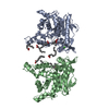
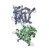
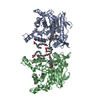
| Title | Staphylococcus aureus lipase-Penfluridol complex (on the ground) | ||||||
 Components Components | Lipase 2 | ||||||
 Keywords Keywords | HYDROLASE / fatty acid binding | ||||||
| Function / homology |  Function and homology information Function and homology informationtriacylglycerol lipase / triacylglycerol lipase activity / lipid catabolic process / extracellular region / metal ion binding Similarity search - Function | ||||||
| Biological species |   Staphylococcus aureus (bacteria) Staphylococcus aureus (bacteria) | ||||||
| Method |  X-RAY DIFFRACTION / SYNCHROTRON / MOLECULAR REPLACEMENT / Resolution: 2.23 Å X-RAY DIFFRACTION / SYNCHROTRON / MOLECULAR REPLACEMENT / Resolution: 2.23 Å | ||||||
 Authors Authors | Kitadokoro, J. / Hirokawa, T. / Kamitani, S. / Kitadokoro, K. | ||||||
| Funding support |  Japan, 1items Japan, 1items
| ||||||
 Citation Citation | Journal: Sci Rep / Year: 2025 Title: Structural analysis shows the mode of inhibition for Staphylococcus aureus lipase by antipsychotic penfluridol. Authors: Kitadokoro, J. / Hirokawa, T. / Kamo, M. / Furubayashi, N. / Okuno, Y. / Hikima, T. / Yamamoto, M. / Inaka, K. / Maenaka, K. / Kamitani, S. / Kitadokoro, K. | ||||||
| History |
|
- Structure visualization
| Structure viewer | Molecule: MolmilJmol/JSmol |
|---|
- Downloads & links
-Download
| PDBx/mmCIF format | 9l3c.cif.gz | 337.9 KB | Display | PDBx/mmCIF format |
|---|---|---|---|---|
| PDB format | pdb9l3c.ent.gz | Display | PDB format | |
| PDBx/mmJSON format | 9l3c.json.gz | Tree view | PDBx/mmJSON format | |
| Others |  Other downloads Other downloads |
-Validation report
| Arichive directory | https://data.pdbj.org/pub/pdb/validation_reports/l3/9l3cftp://data.pdbj.org/pub/pdb/validation_reports/l3/9l3c | HTTPS FTP |
|---|
-Related structure data


-Links
 PDBj
PDBj
- Assembly
| Deposited unit | 
| ||||||||
|---|---|---|---|---|---|---|---|---|---|
| 1 |
| ||||||||
| Unit cell |
| ||||||||
| Components on special symmetry positions |
|
-Components
-Protein , 1 types, 2 molecules AB
| #1: Protein | Mass: 45797.391 Da / Num. of mol.: 2 Source method: isolated from a genetically manipulated source Source: (gene. exp.) Staphylococcus aureus (bacteria) / Gene: lip, BN1321_80040 / Production host: |
|---|
-Non-polymers , 13 types, 79 molecules 
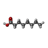
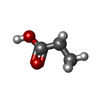


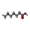


| #2: Chemical |  Mass: 92.094 Da / Num. of mol.: 2 / Source method: obtained synthetically / Formula: C3H8O3 Mass: 92.094 Da / Num. of mol.: 2 / Source method: obtained synthetically / Formula: C3H8O3#3: Chemical |  Mass: 144.211 Da / Num. of mol.: 3 / Source method: obtained synthetically / Formula: C8H16O2 Mass: 144.211 Da / Num. of mol.: 3 / Source method: obtained synthetically / Formula: C8H16O2#4: Chemical | ChemComp-PPI /  Mass: 74.079 Da / Num. of mol.: 5 / Source method: obtained synthetically / Formula: C3H6O2 Mass: 74.079 Da / Num. of mol.: 5 / Source method: obtained synthetically / Formula: C3H6O2#5: Chemical | Mass: 523.965 Da / Num. of mol.: 2 / Source method: obtained synthetically / Formula: C28H27ClF5NO #6: Chemical | ChemComp-FMT /  Mass: 46.025 Da / Num. of mol.: 4 / Source method: obtained synthetically / Formula: CH2O2 Mass: 46.025 Da / Num. of mol.: 4 / Source method: obtained synthetically / Formula: CH2O2#7: Chemical | ChemComp-BUA /  Mass: 88.105 Da / Num. of mol.: 4 / Source method: obtained synthetically / Formula: C4H8O2 Mass: 88.105 Da / Num. of mol.: 4 / Source method: obtained synthetically / Formula: C4H8O2#8: Chemical |  Mass: 65.409 Da / Num. of mol.: 2 / Source method: obtained synthetically / Formula: Zn Mass: 65.409 Da / Num. of mol.: 2 / Source method: obtained synthetically / Formula: Zn#9: Chemical |  Mass: 40.078 Da / Num. of mol.: 2 / Source method: obtained synthetically / Formula: Ca Mass: 40.078 Da / Num. of mol.: 2 / Source method: obtained synthetically / Formula: Ca#10: Chemical | ChemComp-CL /  Mass: 35.453 Da / Num. of mol.: 4 / Source method: obtained synthetically / Formula: Cl Mass: 35.453 Da / Num. of mol.: 4 / Source method: obtained synthetically / Formula: Cl#11: Chemical |  Mass: 186.291 Da / Num. of mol.: 2 / Source method: obtained synthetically / Formula: C11H22O2 / Comment: antifungal*YM Mass: 186.291 Da / Num. of mol.: 2 / Source method: obtained synthetically / Formula: C11H22O2 / Comment: antifungal*YM#12: Chemical | ChemComp-SHV / |  Mass: 130.185 Da / Num. of mol.: 1 / Source method: isolated from a natural source / Formula: C7H14O2 Mass: 130.185 Da / Num. of mol.: 1 / Source method: isolated from a natural source / Formula: C7H14O2#13: Chemical |  Mass: 116.158 Da / Num. of mol.: 2 / Source method: obtained synthetically / Formula: C6H12O2 Mass: 116.158 Da / Num. of mol.: 2 / Source method: obtained synthetically / Formula: C6H12O2#14: Water | ChemComp-HOH / | Mass: 18.015 Da / Num. of mol.: 46 / Source method: isolated from a natural source / Formula: H2O |
|---|
-Details
| Has ligand of interest | N |
|---|---|
| Has protein modification | N |
-Experimental details
-Experiment
| Experiment | Method: X-RAY DIFFRACTION / Number of used crystals: 1 |
|---|
- Sample preparation
| Crystal | Density Matthews: 5.94 Å3/Da / Density % sol: 79.3 % / Description: hexagonal diamond shape |
|---|---|
| Crystal grow | Temperature: 295 K / Method: vapor diffusion, sitting drop / pH: 6.5 / Details: 2.8M Sodium Formate, Acetate |
-Data collection
| Diffraction | Mean temperature: 100 K / Serial crystal experiment: N |
|---|---|
| Diffraction source | Source: SYNCHROTRON / Site: SPring-8 / Beamline: BL41XU / Wavelength: 0.9 Å |
| Detector | Type: DECTRIS EIGER X 16M / Detector: PIXEL / Date: Jun 21, 2023 |
| Radiation | Protocol: SINGLE WAVELENGTH / Monochromatic (M) / Laue (L): M / Scattering type: x-ray |
| Radiation wavelength | Wavelength: 0.9 Å / Relative weight: 1 |
| Reflection | Resolution: 2.23→50 Å / Num. obs: 192595 / % possible obs: 99.8 % / Redundancy: 13.1 % / CC1/2: 0.998 / Net I/σ(I): 12.4 |
| Reflection shell | Resolution: 2.23→2.37 Å / Redundancy: 13 % / Num. unique obs: 7034 / CC1/2: 0.457 / % possible all: 98.7 |
- Processing
| Software |
| ||||||||||||||||||||||||||||||||||||||||||||||||||||||||||||||||||||||||||||||||||||||||||||||||||||||||||||||||||||||||||||||||||||||||||||||||||||||||||||||||||||||||||||||||||||||
|---|---|---|---|---|---|---|---|---|---|---|---|---|---|---|---|---|---|---|---|---|---|---|---|---|---|---|---|---|---|---|---|---|---|---|---|---|---|---|---|---|---|---|---|---|---|---|---|---|---|---|---|---|---|---|---|---|---|---|---|---|---|---|---|---|---|---|---|---|---|---|---|---|---|---|---|---|---|---|---|---|---|---|---|---|---|---|---|---|---|---|---|---|---|---|---|---|---|---|---|---|---|---|---|---|---|---|---|---|---|---|---|---|---|---|---|---|---|---|---|---|---|---|---|---|---|---|---|---|---|---|---|---|---|---|---|---|---|---|---|---|---|---|---|---|---|---|---|---|---|---|---|---|---|---|---|---|---|---|---|---|---|---|---|---|---|---|---|---|---|---|---|---|---|---|---|---|---|---|---|---|---|---|---|
| Refinement | Method to determine structure: MOLECULAR REPLACEMENT / Resolution: 2.23→47 Å / Cor.coef. Fo:Fc: 0.967 / Cor.coef. Fo:Fc free: 0.957 / SU B: 13.954 / SU ML: 0.136 / Cross valid method: THROUGHOUT / ESU R: 0.174 / ESU R Free: 0.131 / Stereochemistry target values: MAXIMUM LIKELIHOOD / Details: HYDROGENS HAVE BEEN ADDED IN THE RIDING POSITIONS
| ||||||||||||||||||||||||||||||||||||||||||||||||||||||||||||||||||||||||||||||||||||||||||||||||||||||||||||||||||||||||||||||||||||||||||||||||||||||||||||||||||||||||||||||||||||||
| Solvent computation | Ion probe radii: 0.8 Å / Shrinkage radii: 0.8 Å / VDW probe radii: 1.2 Å / Solvent model: MASK | ||||||||||||||||||||||||||||||||||||||||||||||||||||||||||||||||||||||||||||||||||||||||||||||||||||||||||||||||||||||||||||||||||||||||||||||||||||||||||||||||||||||||||||||||||||||
| Displacement parameters | Biso mean: 78.31 Å2
| ||||||||||||||||||||||||||||||||||||||||||||||||||||||||||||||||||||||||||||||||||||||||||||||||||||||||||||||||||||||||||||||||||||||||||||||||||||||||||||||||||||||||||||||||||||||
| Refinement step | Cycle: 1 / Resolution: 2.23→47 Å
| ||||||||||||||||||||||||||||||||||||||||||||||||||||||||||||||||||||||||||||||||||||||||||||||||||||||||||||||||||||||||||||||||||||||||||||||||||||||||||||||||||||||||||||||||||||||
| Refine LS restraints |
|