+ Open data
- Basic information
| Entry | Database: PDB / ID: 9l2e | |||||||||||||||||||||
|---|---|---|---|---|---|---|---|---|---|---|---|---|---|---|---|---|---|---|---|---|---|---|
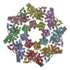
| Title | Structure of SARM1 bound to M1 in the intermediate state 2 | |||||||||||||||||||||
 Components Components | NAD(+) hydrolase SARM1 | |||||||||||||||||||||
 Keywords Keywords | HYDROLASE / NAD(+) hydrolase / cell death / apoptosis | |||||||||||||||||||||
| Function / homology |  Function and homology information Function and homology informationnegative regulation of MyD88-independent toll-like receptor signaling pathway / extrinsic component of synaptic membrane / MyD88-independent TLR4 cascade / NADP+ nucleosidase activity / Toll Like Receptor 3 (TLR3) Cascade / NAD+ catabolic process / NAD+ nucleosidase activity / regulation of synapse pruning / modification of postsynaptic structure / ADP-ribosyl cyclase/cyclic ADP-ribose hydrolase ...negative regulation of MyD88-independent toll-like receptor signaling pathway / extrinsic component of synaptic membrane / MyD88-independent TLR4 cascade / NADP+ nucleosidase activity / Toll Like Receptor 3 (TLR3) Cascade / NAD+ catabolic process / NAD+ nucleosidase activity / regulation of synapse pruning / modification of postsynaptic structure / ADP-ribosyl cyclase/cyclic ADP-ribose hydrolase / protein localization to mitochondrion / NAD+ nucleosidase activity, cyclic ADP-ribose generating / nervous system process / Hydrolases; Glycosylases; Hydrolysing N-glycosyl compounds / regulation of dendrite morphogenesis / response to glucose / response to axon injury / signaling adaptor activity / TRAF6-mediated induction of TAK1 complex within TLR4 complex / regulation of neuron apoptotic process / Activation of IRF3, IRF7 mediated by TBK1, IKKε (IKBKE) / IKK complex recruitment mediated by RIP1 / neuromuscular junction / nervous system development / microtubule / mitochondrial outer membrane / cell differentiation / axon / innate immune response / synapse / dendrite / glutamatergic synapse / cell surface / signal transduction / protein-containing complex / mitochondrion / identical protein binding / cytoplasm / cytosol Similarity search - Function | |||||||||||||||||||||
| Biological species |  Homo sapiens (human) Homo sapiens (human) | |||||||||||||||||||||
| Method | ELECTRON MICROSCOPY / single particle reconstruction / cryo EM / Resolution: 2.46 Å | |||||||||||||||||||||
 Authors Authors | Huang, Y. / Zhang, J. / Zheng, S. / Wang, X. | |||||||||||||||||||||
| Funding support |  China, 1items China, 1items
| |||||||||||||||||||||
 Citation Citation |  Journal: Proc Natl Acad Sci U S A / Year: 2025 Journal: Proc Natl Acad Sci U S A / Year: 2025Title: Stepwise activation of SARM1 for cell death and axon degeneration revealed by a biosynthetic NMN mimic. Authors: Yinpin Huang / Jun Zhang / Wenbin Zhang / Jie Chen / Sijia Chen / Qincui Wu / Sanduo Zheng / Xiaodong Wang /  Abstract: Axon degeneration, driven by the NAD hydrolyzing enzyme SARM1, is an early pathological hallmark of numerous neurodegenerative diseases. SARM1 exists in an inactive form and is activated following ...Axon degeneration, driven by the NAD hydrolyzing enzyme SARM1, is an early pathological hallmark of numerous neurodegenerative diseases. SARM1 exists in an inactive form and is activated following nerve injury. However, the precise molecular mechanism underlying SARM1 activation remains to be fully elucidated. In this study, we report the identification of a potent proactivator of SARM1, G10, which is converted into a direct activator (M1) by the enzyme nicotinamide phosphoribosyltransferase. Cryoelectron microscopy structures of SARM1 bound to M1, as well as to M1 and a nonhydrolyzable NAD analog (1AD), captured two intermediate activation states and the fully active state, revealing a stepwise mechanism of SARM1 activation. Further, introducing a disulfide bond to prevent conformational transitions between the two intermediate states mediated by M1 stabilized SARM1 in its inactive form and blocked M1-induced cell death. Together, these findings propose a sequential, stepwise activation model for SARM1 and offer a framework for developing potential SARM1 inhibitors for the treatment of neurodegenerative diseases. | |||||||||||||||||||||
| History |
|
- Structure visualization
| Structure viewer | Molecule:  Molmil Molmil Jmol/JSmol Jmol/JSmol |
|---|
- Downloads & links
- Download
| PDBx/mmCIF format |  9l2e.cif.gz 9l2e.cif.gz | 261.7 KB | Display |  PDBx/mmCIF format PDBx/mmCIF format |
|---|---|---|---|---|
| PDB format |  pdb9l2e.ent.gz pdb9l2e.ent.gz | 181.6 KB | Display |  PDB format PDB format |
| PDBx/mmJSON format |  9l2e.json.gz 9l2e.json.gz | Tree view |  PDBx/mmJSON format PDBx/mmJSON format | |
| Others |  Other downloads Other downloads |
-Validation report
| Arichive directory |  https://data.pdbj.org/pub/pdb/validation_reports/l2/9l2e https://data.pdbj.org/pub/pdb/validation_reports/l2/9l2e ftp://data.pdbj.org/pub/pdb/validation_reports/l2/9l2e ftp://data.pdbj.org/pub/pdb/validation_reports/l2/9l2e | HTTPS FTP |
|---|
-Related structure data
| Related structure data |  62773MC  9l2dC  9l2fC  9l2gC C: citing same article ( M: map data used to model this data |
|---|---|
| Similar structure data | Similarity search - Function & homology  F&H Search F&H Search |
- Links
Links
- Assembly
| Deposited unit | 
|
|---|---|
| 1 |
|
- Components
| #1: Protein | Mass: 80483.133 Da / Num. of mol.: 8 Source method: isolated from a genetically manipulated source Source: (gene. exp.)  Homo sapiens (human) / Gene: SARM1, KIAA0524, SAMD2, SARM / Production host: Homo sapiens (human) / Gene: SARM1, KIAA0524, SAMD2, SARM / Production host:  Homo sapiens (human) Homo sapiens (human)References: UniProt: Q6SZW1, ADP-ribosyl cyclase/cyclic ADP-ribose hydrolase, Hydrolases; Glycosylases; Hydrolysing N-glycosyl compounds Has protein modification | N | |
|---|
-Experimental details
-Experiment
| Experiment | Method: ELECTRON MICROSCOPY |
|---|---|
| EM experiment | Aggregation state: PARTICLE / 3D reconstruction method: single particle reconstruction |
- Sample preparation
| Component | Name: Structure of SARM1 bound to M1 in the intermediate state 2 Type: COMPLEX / Entity ID: all / Source: RECOMBINANT |
|---|---|
| Source (natural) | Organism:  Homo sapiens (human) Homo sapiens (human) |
| Source (recombinant) | Organism:  Homo sapiens (human) / Strain: HEK293T Homo sapiens (human) / Strain: HEK293T |
| Buffer solution | pH: 7.4 |
| Specimen | Embedding applied: NO / Shadowing applied: NO / Staining applied: NO / Vitrification applied: YES |
| Vitrification | Cryogen name: ETHANE |
- Electron microscopy imaging
| Experimental equipment |  Model: Titan Krios / Image courtesy: FEI Company |
|---|---|
| Microscopy | Model: TFS KRIOS |
| Electron gun | Electron source:  FIELD EMISSION GUN / Accelerating voltage: 300 kV / Illumination mode: FLOOD BEAM FIELD EMISSION GUN / Accelerating voltage: 300 kV / Illumination mode: FLOOD BEAM |
| Electron lens | Mode: BRIGHT FIELD / Nominal defocus max: 2240 nm / Nominal defocus min: 710 nm |
| Image recording | Electron dose: 52.73 e/Å2 / Film or detector model: GATAN K3 BIOQUANTUM (6k x 4k) |
- Processing
| EM software | Name: PHENIX / Version: 1.19_4092 / Category: model refinement | ||||||||||||||||||||||||
|---|---|---|---|---|---|---|---|---|---|---|---|---|---|---|---|---|---|---|---|---|---|---|---|---|---|
| CTF correction | Type: PHASE FLIPPING AND AMPLITUDE CORRECTION | ||||||||||||||||||||||||
| 3D reconstruction | Resolution: 2.46 Å / Resolution method: FSC 0.143 CUT-OFF / Num. of particles: 244752 / Symmetry type: POINT | ||||||||||||||||||||||||
| Refinement | Highest resolution: 2.46 Å Stereochemistry target values: REAL-SPACE (WEIGHTED MAP SUM AT ATOM CENTERS) | ||||||||||||||||||||||||
| Refine LS restraints |
|
 Movie
Movie Controller


 PDBj
PDBj


