 Movie
Movie Controller

[English] 日本語
 Yorodumi
- PDB-9l04: Crystal structure of human ALK2 kinase domain with R206H mutation... -
+ Open data

- Basic information
| Entry | Database: PDB / ID: 9l04 | ||||||
|---|---|---|---|---|---|---|---|
| Title | Crystal structure of human ALK2 kinase domain with R206H mutation in complex with RK783 | ||||||
 Components Components | Activin receptor type-1 | ||||||
 Keywords Keywords | SIGNALING PROTEIN / KINASE / INHIBITOR COMPLEX | ||||||
| Function / homology |  Function and homology information Function and homology informationendocardial cushion cell fate commitment / mitral valve morphogenesis / BMP receptor complex / cardiac muscle cell fate commitment / BMP receptor activity / endocardial cushion fusion / atrial septum primum morphogenesis / positive regulation of cardiac epithelial to mesenchymal transition / acute inflammatory response / positive regulation of determination of dorsal identity ...endocardial cushion cell fate commitment / mitral valve morphogenesis / BMP receptor complex / cardiac muscle cell fate commitment / BMP receptor activity / endocardial cushion fusion / atrial septum primum morphogenesis / positive regulation of cardiac epithelial to mesenchymal transition / acute inflammatory response / positive regulation of determination of dorsal identity / transforming growth factor beta receptor activity, type I / smooth muscle cell differentiation / activin receptor complex / activin receptor activity, type I / endocardial cushion formation / pharyngeal system development / transmembrane receptor protein serine/threonine kinase activity / receptor protein serine/threonine kinase / activin binding / cellular response to BMP stimulus / activin receptor signaling pathway / negative regulation of activin receptor signaling pathway / embryonic heart tube morphogenesis / gastrulation with mouth forming second / dorsal/ventral pattern formation / transforming growth factor beta binding / determination of left/right symmetry / atrioventricular valve morphogenesis / neural crest cell migration / branching involved in blood vessel morphogenesis / negative regulation of G1/S transition of mitotic cell cycle / ventricular septum morphogenesis / SMAD binding / germ cell development / peptide hormone binding / mesoderm formation / positive regulation of intracellular signal transduction / positive regulation of SMAD protein signal transduction / regulation of ossification / positive regulation of bone mineralization / positive regulation of osteoblast differentiation / BMP signaling pathway / negative regulation of signal transduction / transforming growth factor beta receptor signaling pathway / protein tyrosine kinase binding / negative regulation of extrinsic apoptotic signaling pathway / cellular response to growth factor stimulus / apical part of cell / osteoblast differentiation / heart development / in utero embryonic development / cell differentiation / protein kinase activity / positive regulation of cell migration / cadherin binding / protein serine/threonine kinase activity / positive regulation of DNA-templated transcription / protein homodimerization activity / positive regulation of transcription by RNA polymerase II / ATP binding / metal ion binding / plasma membrane Similarity search - Function | ||||||
| Biological species |  Homo sapiens (human) Homo sapiens (human) | ||||||
| Method |  X-RAY DIFFRACTION / SYNCHROTRON / MOLECULAR REPLACEMENT / Resolution: 2.25 Å X-RAY DIFFRACTION / SYNCHROTRON / MOLECULAR REPLACEMENT / Resolution: 2.25 Å | ||||||
 Authors Authors | Sakai, N. / Mishima-Tsumagari, C. / Shirouzu, M. | ||||||
| Funding support | 1items
| ||||||
 Citation Citation | Journal: Bone / Year: 2025 Title: A new BMP type 1 receptor kinase inhibitor for safe and efficient oral treatment to prevent genetically induced heterotopic ossification in mice. Authors: Yang, J. / Pan, H. / Sekimata, K. / Hwang, C. / Kulkarni, A. / Thomas, H. / Lindenau, J. / Duford, T. / Ueharu, H. / Tanaka, A. / Sakai, N. / Shirouzu, M. / Hashizume, Y. / Levi, B. / Koyama, H. / Mishina, Y. | ||||||
| History |
|
- Structure visualization
| Structure viewer | Molecule: MolmilJmol/JSmol |
|---|
- Downloads & links
-Download
| PDBx/mmCIF format | 9l04.cif.gz | 94.5 KB | Display | PDBx/mmCIF format |
|---|---|---|---|---|
| PDB format | pdb9l04.ent.gz | Display | PDB format | |
| PDBx/mmJSON format | 9l04.json.gz | Tree view | PDBx/mmJSON format | |
| Others |  Other downloads Other downloads |
-Validation report
| Summary document | 9l04_validation.pdf.gz | 719.7 KB | Display | wwPDB validaton report |
|---|---|---|---|---|
| Full document | 9l04_full_validation.pdf.gz | 724.1 KB | Display | |
| Data in XML | 9l04_validation.xml.gz | 16.6 KB | Display | |
| Data in CIF | 9l04_validation.cif.gz | 22.6 KB | Display | |
| Arichive directory | https://data.pdbj.org/pub/pdb/validation_reports/l0/9l04ftp://data.pdbj.org/pub/pdb/validation_reports/l0/9l04 | HTTPS FTP |
-Related structure data
| Related structure data | 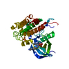 6juxS S: Starting model for refinement |
|---|---|
| Similar structure data |
-Links
 PDBj
PDBj
- Assembly
| Deposited unit | 
| ||||||||||||
|---|---|---|---|---|---|---|---|---|---|---|---|---|---|
| 1 |
| ||||||||||||
| Unit cell |
|
-Components
| #1: Protein | Mass: 34531.625 Da / Num. of mol.: 1 / Mutation: R206H Source method: isolated from a genetically manipulated source Details: N-TERMINAL 2 RESIDUES "SM" ARE THE RESIDUAL OF THE PURIFICATION TAG AFTER CLEAVING OFF THE TAG BY PROTEASE, FOLLOWED BY THE Q201 OF ALK2 Source: (gene. exp.) Homo sapiens (human) / Gene: ACVR1, ACVRLK2 / Production host:   Spodoptera frugiperda (fall armyworm) Spodoptera frugiperda (fall armyworm)References: UniProt: Q04771, receptor protein serine/threonine kinase | ||||||
|---|---|---|---|---|---|---|---|
| #2: Chemical | ChemComp-A1L4C / Mass: 482.580 Da / Num. of mol.: 1 / Source method: obtained synthetically / Formula: C27H30N8O / Feature type: SUBJECT OF INVESTIGATION | ||||||
| #3: Chemical | ChemComp-SO4 /   Mass: 96.063 Da / Num. of mol.: 6 / Source method: obtained synthetically / Formula: SO4 Mass: 96.063 Da / Num. of mol.: 6 / Source method: obtained synthetically / Formula: SO4#4: Water | ChemComp-HOH / |  Mass: 18.015 Da / Num. of mol.: 112 / Source method: isolated from a natural source / Formula: H2O Mass: 18.015 Da / Num. of mol.: 112 / Source method: isolated from a natural source / Formula: H2OHas ligand of interest | Y | Has protein modification | N | |
-Experimental details
-Experiment
| Experiment | Method: X-RAY DIFFRACTION / Number of used crystals: 1 |
|---|
- Sample preparation
| Crystal | Density Matthews: 2.59 Å3/Da / Density % sol: 52.46 % |
|---|---|
| Crystal grow | Temperature: 293 K / Method: vapor diffusion, sitting drop / Details: 100 mM HEPES, 1.5 -1.6 M Ammonium sulfate / PH range: 7.6 - 8.0 |
-Data collection
| Diffraction | Mean temperature: 100 K / Serial crystal experiment: N | |||||||||||||||||||||||||||||||||||||||||||||||||||||||||||||||||||||||||||||||||||||||||||||||||||
|---|---|---|---|---|---|---|---|---|---|---|---|---|---|---|---|---|---|---|---|---|---|---|---|---|---|---|---|---|---|---|---|---|---|---|---|---|---|---|---|---|---|---|---|---|---|---|---|---|---|---|---|---|---|---|---|---|---|---|---|---|---|---|---|---|---|---|---|---|---|---|---|---|---|---|---|---|---|---|---|---|---|---|---|---|---|---|---|---|---|---|---|---|---|---|---|---|---|---|---|---|
| Diffraction source | Source: SYNCHROTRON / Site: SPring-8  / Beamline: BL26B2 / Wavelength: 1 Å / Beamline: BL26B2 / Wavelength: 1 Å | |||||||||||||||||||||||||||||||||||||||||||||||||||||||||||||||||||||||||||||||||||||||||||||||||||
| Detector | Type: RAYONIX MX225-HS / Detector: CCD / Date: Apr 13, 2017 | |||||||||||||||||||||||||||||||||||||||||||||||||||||||||||||||||||||||||||||||||||||||||||||||||||
| Radiation | Monochromator: Silicon double crystals / Protocol: SINGLE WAVELENGTH / Monochromatic (M) / Laue (L): M / Scattering type: x-ray | |||||||||||||||||||||||||||||||||||||||||||||||||||||||||||||||||||||||||||||||||||||||||||||||||||
| Radiation wavelength | Wavelength: 1 Å / Relative weight: 1 | |||||||||||||||||||||||||||||||||||||||||||||||||||||||||||||||||||||||||||||||||||||||||||||||||||
| Reflection | Resolution: 2.25→46.05 Å / Num. obs: 32655 / % possible obs: 99.46 % / Redundancy: 7.31 % / Biso Wilson estimate: 35.65 Å2 / CC1/2: 0.999 / Rmerge(I) obs: 0.067 / Rpim(I) all: 0.026 / Rrim(I) all: 0.072 / Net I/σ(I): 18.6 | |||||||||||||||||||||||||||||||||||||||||||||||||||||||||||||||||||||||||||||||||||||||||||||||||||
| Reflection shell | Diffraction-ID: 1
|
- Processing
| Software |
| |||||||||||||||||||||||||||||||||||||||||||||||||||||||||||||||||||||||||||||||||||||||||||
|---|---|---|---|---|---|---|---|---|---|---|---|---|---|---|---|---|---|---|---|---|---|---|---|---|---|---|---|---|---|---|---|---|---|---|---|---|---|---|---|---|---|---|---|---|---|---|---|---|---|---|---|---|---|---|---|---|---|---|---|---|---|---|---|---|---|---|---|---|---|---|---|---|---|---|---|---|---|---|---|---|---|---|---|---|---|---|---|---|---|---|---|---|
| Refinement | Method to determine structure: MOLECULAR REPLACEMENT Starting model: 6JUX Resolution: 2.25→46.04 Å / SU ML: 0.2955 / Cross valid method: FREE R-VALUE / σ(F): 1.38 / Phase error: 28.4696 Stereochemistry target values: GeoStd + Monomer Library + CDL v1.2
| |||||||||||||||||||||||||||||||||||||||||||||||||||||||||||||||||||||||||||||||||||||||||||
| Solvent computation | Shrinkage radii: 0.9 Å / VDW probe radii: 1.1 Å / Solvent model: FLAT BULK SOLVENT MODEL | |||||||||||||||||||||||||||||||||||||||||||||||||||||||||||||||||||||||||||||||||||||||||||
| Displacement parameters | Biso mean: 41.72 Å2 | |||||||||||||||||||||||||||||||||||||||||||||||||||||||||||||||||||||||||||||||||||||||||||
| Refinement step | Cycle: LAST / Resolution: 2.25→46.04 Å
| |||||||||||||||||||||||||||||||||||||||||||||||||||||||||||||||||||||||||||||||||||||||||||
| Refine LS restraints |
| |||||||||||||||||||||||||||||||||||||||||||||||||||||||||||||||||||||||||||||||||||||||||||
| LS refinement shell |
|
