Movie
Movie Controller

[English] 日本語
 Yorodumi
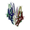
- PDB-9k29: Structure of the Salmonella flagellar FliPQR complex reconstitute... -
+ Open data

- Basic information
| Entry | Database: PDB / ID: 9k29 | |||||||||||||||||||||||||||||||||
|---|---|---|---|---|---|---|---|---|---|---|---|---|---|---|---|---|---|---|---|---|---|---|---|---|---|---|---|---|---|---|---|---|---|---|
| Title | Structure of the Salmonella flagellar FliPQR complex reconstituted in the peptidisc | |||||||||||||||||||||||||||||||||
 Components Components |
| |||||||||||||||||||||||||||||||||
 Keywords Keywords | PROTEIN TRANSPORT / Bacterial flagellum / flagellar assembly / electron Cryomicroscopy / type III secretion system / Salmonella / MOTOR PROTEIN | |||||||||||||||||||||||||||||||||
| Function / homology |  Function and homology information Function and homology informationbacterial-type flagellum basal body / bacterial-type flagellum-dependent swarming motility / bacterial-type flagellum assembly / protein secretion / protein targeting / plasma membrane Similarity search - Function | |||||||||||||||||||||||||||||||||
| Biological species |  Salmonella enterica subsp. enterica serovar Typhimurium str. LT2 (bacteria) Salmonella enterica subsp. enterica serovar Typhimurium str. LT2 (bacteria) | |||||||||||||||||||||||||||||||||
| Method | ELECTRON MICROSCOPY / single particle reconstruction / cryo EM / Resolution: 3 Å | |||||||||||||||||||||||||||||||||
 Authors Authors | Kinoshita, M. / Miyata, T. / Makino, F. / Imada, K. / Namba, K. / Minamino, T. | |||||||||||||||||||||||||||||||||
| Funding support |  Japan, 10items Japan, 10items
| |||||||||||||||||||||||||||||||||
 Citation Citation | Journal: Proc Natl Acad Sci U S A / Year: 2025 Title: A β-cap on the FliPQR protein-export channel acts as the cap for initial flagellar rod assembly. Authors: Miki Kinoshita / Tomoko Miyata / Fumiaki Makino / Katsumi Imada / Keiichi Namba / Tohru Minamino / Abstract: The FliPQR complex constitutes a channel for export of the flagellar proteins involved in axial structure assembly. It also serves as a template for the assembly of the rod structure, which consists ...The FliPQR complex constitutes a channel for export of the flagellar proteins involved in axial structure assembly. It also serves as a template for the assembly of the rod structure, which consists of FliE, FlgB, FlgC, FlgF, and FlgG. FliP, FliQ, and FliR assemble into a right-handed helical structure within the central pore of the flagellar basal body MS-ring, and the complex has two gates on the cytoplasmic and periplasmic sides. The periplasmic gate, formed by the N-terminal α-helices of FliP and FliR, remains closed until six FliE subunits assemble onto FliP and FliR to form the first layer of the rod, but it has remained unclear how each FliE subunit opens the gate and assembles in the absence of the rod cap required for efficient assembly of other rod proteins. Here, we present a cryoelectron microscopy structure of the FliPQR complex in closed form at 3.0 Å resolution. A β-cap, formed by the N-terminal β-strands of FliP and FliR, is located at the top of the FliPQR complex and tightly seals the closed gate. The β-cap has a narrow pore that efficiently and accurately leads the first FliE subunit to its assembly site. Interactions of FliE with FliP and FliR induce a conformational change in FliP and FliR, with their N-terminal α-helices move up and outward to open the gate. Consequently, each of the N-terminal β-strands of FliP and FliR detaches from the β-cap one after another, thereby creating a docking site for the next FliE subunit to efficiently assemble. | |||||||||||||||||||||||||||||||||
| History |
|
- Structure visualization
| Structure viewer | Molecule: MolmilJmol/JSmol |
|---|
- Downloads & links
-Download
| PDBx/mmCIF format | 9k29.cif.gz | 291 KB | Display | PDBx/mmCIF format |
|---|---|---|---|---|
| PDB format | pdb9k29.ent.gz | 236.8 KB | Display | PDB format |
| PDBx/mmJSON format | 9k29.json.gz | Tree view | PDBx/mmJSON format | |
| Others |  Other downloads Other downloads |
-Validation report
| Arichive directory | https://data.pdbj.org/pub/pdb/validation_reports/k2/9k29ftp://data.pdbj.org/pub/pdb/validation_reports/k2/9k29 | HTTPS FTP |
|---|
-Related structure data
| Related structure data |  61993MC M: map data used to model this data C: citing same article ( |
|---|---|
| Similar structure data |
-Links
 PDBj
PDBj
- Assembly
| Deposited unit | 
|
|---|---|
| 1 |
|
-Components
| #1: Protein | Mass: 26801.086 Da / Num. of mol.: 5 Source method: isolated from a genetically manipulated source Source: (gene. exp.) Salmonella enterica subsp. enterica serovar Typhimurium str. LT2 (bacteria)Gene: fliP, flaR, STM1979 Production host: Salmonella enterica subsp. enterica serovar Typhimurium str. LT2 (bacteria)References: UniProt: P54700 #2: Protein | | Mass: 30320.314 Da / Num. of mol.: 1 Source method: isolated from a genetically manipulated source Source: (gene. exp.) Salmonella enterica subsp. enterica serovar Typhimurium str. LT2 (bacteria)Gene: fliR, flaP, STM1981 Production host: Salmonella enterica subsp. enterica serovar Typhimurium str. LT2 (bacteria)References: UniProt: P54702 #3: Protein | Mass: 9606.758 Da / Num. of mol.: 4 Source method: isolated from a genetically manipulated source Source: (gene. exp.) Salmonella enterica subsp. enterica serovar Typhimurium str. LT2 (bacteria)Gene: fliQ, flaQ, STM1980 Production host: Salmonella enterica subsp. enterica serovar Typhimurium str. LT2 (bacteria)References: UniProt: P0A1L5 Has protein modification | N | |
|---|
-Experimental details
-Experiment
| Experiment | Method: ELECTRON MICROSCOPY |
|---|---|
| EM experiment | Aggregation state: PARTICLE / 3D reconstruction method: single particle reconstruction |
- Sample preparation
| Component | Name: Structure of the Salmonella flagellar FliPQR complex reconstituted in the peptidisc Type: COMPLEX / Entity ID: all / Source: RECOMBINANT | |||||||||||||||
|---|---|---|---|---|---|---|---|---|---|---|---|---|---|---|---|---|
| Molecular weight | Experimental value: NO | |||||||||||||||
| Source (natural) | Organism: Salmonella enterica subsp. enterica serovar Typhimurium str. LT2 (bacteria) | |||||||||||||||
| Source (recombinant) | Organism: Salmonella enterica subsp. enterica serovar Typhimurium str. LT2 (bacteria) | |||||||||||||||
| Buffer solution | pH: 8 | |||||||||||||||
| Buffer component |
| |||||||||||||||
| Specimen | Embedding applied: NO / Shadowing applied: NO / Staining applied: NO / Vitrification applied: YES | |||||||||||||||
| Specimen support | Grid material: COPPER / Grid mesh size: 200 divisions/in. / Grid type: Quantifoil R1.2/1.3 | |||||||||||||||
| Vitrification | Instrument: FEI VITROBOT MARK III / Cryogen name: ETHANE / Humidity: 100 % / Chamber temperature: 277 K |
- Electron microscopy imaging
| Microscopy | Model: JEOL CRYO ARM 300 |
|---|---|
| Electron gun | Electron source:  FIELD EMISSION GUN / Accelerating voltage: 300 kV / Illumination mode: FLOOD BEAM FIELD EMISSION GUN / Accelerating voltage: 300 kV / Illumination mode: FLOOD BEAM |
| Electron lens | Mode: BRIGHT FIELD / Nominal magnification: 50000 X / Nominal defocus max: 2500 nm / Nominal defocus min: 500 nm / Cs: 2.7 mm / Alignment procedure: COMA FREE |
| Specimen holder | Cryogen: NITROGEN / Specimen holder model: JEOL CRYOSPECPORTER |
| Image recording | Electron dose: 40 e/Å2 / Film or detector model: GATAN K3 (6k x 4k) / Num. of real images: 4885 |
| EM imaging optics | Energyfilter name: In-column Omega Filter / Energyfilter slit width: 20 eV |
| Image scans | Sampling size: 5 µm / Width: 5760 / Height: 4092 |
- Processing
| EM software | Name: PHENIX / Version: 1.20.1_4487: / Category: model refinement | ||||||||||||||||||||||||||||
|---|---|---|---|---|---|---|---|---|---|---|---|---|---|---|---|---|---|---|---|---|---|---|---|---|---|---|---|---|---|
| CTF correction | Type: PHASE FLIPPING AND AMPLITUDE CORRECTION | ||||||||||||||||||||||||||||
| Particle selection | Num. of particles selected: 1015741 | ||||||||||||||||||||||||||||
| 3D reconstruction | Resolution: 3 Å / Resolution method: FSC 0.143 CUT-OFF / Num. of particles: 109333 / Symmetry type: POINT | ||||||||||||||||||||||||||||
| Atomic model building |
| ||||||||||||||||||||||||||||
| Atomic model building |
| ||||||||||||||||||||||||||||
| Refine LS restraints |
|