 Movie
Movie Controller

[English] 日本語
 Yorodumi
- PDB-9jzf: Crystal structure of OsSPS3 complexed with zoledronate and isopen... -
+ Open data

- Basic information
| Entry | Database: PDB / ID: 9jzf | ||||||
|---|---|---|---|---|---|---|---|
| Title | Crystal structure of OsSPS3 complexed with zoledronate and isopentenyl diphosphate | ||||||
 Components Components | Probable solanesyl-diphosphate synthase 3, chloroplastic | ||||||
 Keywords Keywords | TRANSFERASE / inhibitor / chloroplast | ||||||
| Function / homology |  Function and homology information Function and homology informationall-trans-nonaprenyl diphosphate synthase [geranyl-diphosphate specific] / all-trans-nonaprenyl-diphosphate synthase (geranyl-diphosphate specific) activity / plastoquinone biosynthetic process / prenyltransferase activity / isoprenoid biosynthetic process / chloroplast / metal ion binding Similarity search - Function | ||||||
| Biological species |  | ||||||
| Method |  X-RAY DIFFRACTION / SYNCHROTRON / MOLECULAR REPLACEMENT / Resolution: 2.446 Å X-RAY DIFFRACTION / SYNCHROTRON / MOLECULAR REPLACEMENT / Resolution: 2.446 Å | ||||||
 Authors Authors | Xiao, H. / Li, M. / Yang, G.-F. | ||||||
| Funding support |  China, 1items China, 1items
| ||||||
 Citation Citation | Journal: To Be Published Title: Crystal structure of OsSPS3 complexed with zoledronate and isopentenyl diphosphate Authors: Xiao, H. / Li, M. / Yang, G.-F. | ||||||
| History |
|
- Structure visualization
| Structure viewer | Molecule: MolmilJmol/JSmol |
|---|
- Downloads & links
-Download
| PDBx/mmCIF format | 9jzf.cif.gz | 129.2 KB | Display | PDBx/mmCIF format |
|---|---|---|---|---|
| PDB format | pdb9jzf.ent.gz | 98 KB | Display | PDB format |
| PDBx/mmJSON format | 9jzf.json.gz | Tree view | PDBx/mmJSON format | |
| Others |  Other downloads Other downloads |
-Validation report
| Arichive directory | https://data.pdbj.org/pub/pdb/validation_reports/jz/9jzfftp://data.pdbj.org/pub/pdb/validation_reports/jz/9jzf | HTTPS FTP |
|---|
-Related structure data
| Similar structure data |
|---|
-Links
 PDBj
PDBj
- Assembly
| Deposited unit | 
| ||||||||
|---|---|---|---|---|---|---|---|---|---|
| 1 |
| ||||||||
| Unit cell |
|
-Components
| #1: Protein | Mass: 35590.414 Da / Num. of mol.: 2 Source method: isolated from a genetically manipulated source Source: (gene. exp.) Gene: SPS3, Os12g0271700, LOC_Os12g17320, OsJ_1767i3 / Production host:  References: UniProt: Q0INZ4, all-trans-nonaprenyl diphosphate synthase [geranyl-diphosphate specific] #2: Chemical | ChemComp-IPE / | 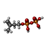  Mass: 246.092 Da / Num. of mol.: 1 / Source method: obtained synthetically / Formula: C5H12O7P2 / Feature type: SUBJECT OF INVESTIGATION Mass: 246.092 Da / Num. of mol.: 1 / Source method: obtained synthetically / Formula: C5H12O7P2 / Feature type: SUBJECT OF INVESTIGATION#3: Chemical | ChemComp-ZOL / |   Mass: 272.090 Da / Num. of mol.: 1 / Source method: obtained synthetically / Formula: C5H10N2O7P2 / Comment: medication*YM Mass: 272.090 Da / Num. of mol.: 1 / Source method: obtained synthetically / Formula: C5H10N2O7P2 / Comment: medication*YM#4: Chemical |   Mass: 24.305 Da / Num. of mol.: 3 / Source method: obtained synthetically / Formula: Mg Mass: 24.305 Da / Num. of mol.: 3 / Source method: obtained synthetically / Formula: Mg#5: Water | ChemComp-HOH / |  Mass: 18.015 Da / Num. of mol.: 23 / Source method: isolated from a natural source / Formula: H2O Mass: 18.015 Da / Num. of mol.: 23 / Source method: isolated from a natural source / Formula: H2OHas ligand of interest | Y | Has protein modification | N | |
|---|
-Experimental details
-Experiment
| Experiment | Method: X-RAY DIFFRACTION / Number of used crystals: 1 |
|---|
- Sample preparation
| Crystal | Density Matthews: 2.41 Å3/Da / Density % sol: 49.02 % |
|---|---|
| Crystal grow | Temperature: 291 K / Method: vapor diffusion, hanging drop / Details: PEG3350, Tris, Ammonium sulfate |
-Data collection
| Diffraction | Mean temperature: 100 K / Serial crystal experiment: N |
|---|---|
| Diffraction source | Source: SYNCHROTRON / Site: SSRF / Beamline: BL10U2 / Wavelength: 0.979183 Å |
| Detector | Type: DECTRIS EIGER X 16M / Detector: PIXEL / Date: Sep 26, 2024 |
| Radiation | Protocol: SINGLE WAVELENGTH / Monochromatic (M) / Laue (L): M / Scattering type: x-ray |
| Radiation wavelength | Wavelength: 0.979183 Å / Relative weight: 1 |
| Reflection | Resolution: 2.446→48.293 Å / Num. obs: 24483 / % possible obs: 98.73 % / Redundancy: 10.244 % / CC1/2: 0.989 / Net I/σ(I): 10.61 |
| Reflection shell | Resolution: 2.446→2.534 Å / Num. unique obs: 2390 / CC1/2: 0.851 |
- Processing
| Software |
| ||||||||||||||||||||||||||||||||||||||||||||||||||||||||||||||||||||||
|---|---|---|---|---|---|---|---|---|---|---|---|---|---|---|---|---|---|---|---|---|---|---|---|---|---|---|---|---|---|---|---|---|---|---|---|---|---|---|---|---|---|---|---|---|---|---|---|---|---|---|---|---|---|---|---|---|---|---|---|---|---|---|---|---|---|---|---|---|---|---|---|
| Refinement | Method to determine structure: MOLECULAR REPLACEMENT / Resolution: 2.446→48.293 Å / SU ML: 0.38 / Cross valid method: FREE R-VALUE / σ(F): 1.34 / Phase error: 36.14 / Stereochemistry target values: ML
| ||||||||||||||||||||||||||||||||||||||||||||||||||||||||||||||||||||||
| Solvent computation | Shrinkage radii: 0.9 Å / VDW probe radii: 1.11 Å / Solvent model: FLAT BULK SOLVENT MODEL | ||||||||||||||||||||||||||||||||||||||||||||||||||||||||||||||||||||||
| Refinement step | Cycle: LAST / Resolution: 2.446→48.293 Å
| ||||||||||||||||||||||||||||||||||||||||||||||||||||||||||||||||||||||
| Refine LS restraints |
| ||||||||||||||||||||||||||||||||||||||||||||||||||||||||||||||||||||||
| LS refinement shell |
|
