 Movie
Movie Controller


+ Open data

- Basic information
| Entry | Database: PDB / ID: 9jtw | ||||||||||||
|---|---|---|---|---|---|---|---|---|---|---|---|---|---|
| Title | AtALMT1 with LMNG and sterol mimic CHS | ||||||||||||
 Components Components | Aluminum-activated malate transporter 1 | ||||||||||||
 Keywords Keywords | MEMBRANE PROTEIN / Plant / Channel | ||||||||||||
| Function / homology | malate transmembrane transport / malate transmembrane transporter activity / Aluminum-activated malate transporter / Aluminium activated malate transporter / response to aluminum ion / monoatomic ion transmembrane transport / plasma membrane / Aluminum-activated malate transporter 1 Function and homology information Function and homology information | ||||||||||||
| Biological species |  | ||||||||||||
| Method | ELECTRON MICROSCOPY / single particle reconstruction / cryo EM / Resolution: 3.7 Å | ||||||||||||
 Authors Authors | Lee, Y. / Lee, S. | ||||||||||||
| Funding support |  Korea, Republic Of, 3items Korea, Republic Of, 3items
| ||||||||||||
 Citation Citation | Journal: Nat Commun / Year: 2025 Title: Structural basis for malate-driven, pore lipid-regulated activation of the Arabidopsis vacuolar anion channel ALMT9. Authors: Yeongmok Lee / Elsa Demes-Causse / Jaemin Yoo / Seo Young Jang / Seoyeon Jung / Justyna Jaślan / Geum-Sook Hwang / Jejoong Yoo / Alexis De Angeli / Sangho Lee /  Abstract: In plant cells, ALMTs are key plasma and vacuolar membrane-localized anion channels regulating plant responses to the environment. Vacuolar ALMTs control anion accumulation in plant cells and, in ...In plant cells, ALMTs are key plasma and vacuolar membrane-localized anion channels regulating plant responses to the environment. Vacuolar ALMTs control anion accumulation in plant cells and, in guard cells, they regulate stomata aperture. The activation of vacuolar ALMTs depends on voltage and cytosolic malate, but the underlying molecular mechanisms remain elusive. Here we report the cryo-EM structures of ALMT9 from Arabidopsis thaliana (AtALMT9), a malate-activated vacuolar anion channel, in plugged and unplugged lipid-bound states. In all these states, membrane lipids interact with the ion conduction pathway of AtALMT9. We identify two unplugged states presenting two distinct pore width profiles. Combining structural and functional analysis we identified conserved residues involved in ion conduction and in the pore lipid interaction. Molecular dynamics simulations revealed a peculiar anion conduction mechanism in AtALMT9. We propose a voltage-dependent activation mechanism based on the competition between pore lipids and malate at the cytosolic entrance of the channel. #1: Journal: Acta Crystallogr D Struct Biol / Year: 2018 Title: Real-space refinement in PHENIX for cryo-EM and crystallography. Authors: Pavel V Afonine / Billy K Poon / Randy J Read / Oleg V Sobolev / Thomas C Terwilliger / Alexandre Urzhumtsev / Paul D Adams /   Abstract: This article describes the implementation of real-space refinement in the phenix.real_space_refine program from the PHENIX suite. The use of a simplified refinement target function enables very fast ...This article describes the implementation of real-space refinement in the phenix.real_space_refine program from the PHENIX suite. The use of a simplified refinement target function enables very fast calculation, which in turn makes it possible to identify optimal data-restraint weights as part of routine refinements with little runtime cost. Refinement of atomic models against low-resolution data benefits from the inclusion of as much additional information as is available. In addition to standard restraints on covalent geometry, phenix.real_space_refine makes use of extra information such as secondary-structure and rotamer-specific restraints, as well as restraints or constraints on internal molecular symmetry. The re-refinement of 385 cryo-EM-derived models available in the Protein Data Bank at resolutions of 6 Å or better shows significant improvement of the models and of the fit of these models to the target maps. | ||||||||||||
| History |
|
- Structure visualization
| Structure viewer | Molecule: MolmilJmol/JSmol |
|---|
- Downloads & links
-Download
| PDBx/mmCIF format | 9jtw.cif.gz | 161.7 KB | Display | PDBx/mmCIF format |
|---|---|---|---|---|
| PDB format | pdb9jtw.ent.gz | 114.7 KB | Display | PDB format |
| PDBx/mmJSON format | 9jtw.json.gz | Tree view | PDBx/mmJSON format | |
| Others |  Other downloads Other downloads |
-Validation report
| Arichive directory | https://data.pdbj.org/pub/pdb/validation_reports/jt/9jtwftp://data.pdbj.org/pub/pdb/validation_reports/jt/9jtw | HTTPS FTP |
|---|
-Related structure data
| Related structure data | 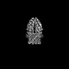 61818MC 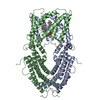 8zteC 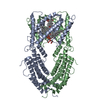 8ztgC 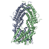 8zthC  8ztiC  8ztjC  8ztkC  8ztlC  8ztmC  8ztnC M: map data used to model this data C: citing same article ( |
|---|---|
| Similar structure data |


-Links
 PDBj
PDBj- Assembly
| Deposited unit | 
|
|---|---|
| 1 |
|
-Components
| #1: Protein | Mass: 55146.891 Da / Num. of mol.: 2 Source method: isolated from a genetically manipulated source Source: (gene. exp.)   Spodoptera frugiperda (fall armyworm) / References: UniProt: Q9SJE9 Spodoptera frugiperda (fall armyworm) / References: UniProt: Q9SJE9Has protein modification | N | |
|---|
-Experimental details
-Experiment
| Experiment | Method: ELECTRON MICROSCOPY |
|---|---|
| EM experiment | Aggregation state: PARTICLE / 3D reconstruction method: single particle reconstruction |
- Sample preparation
| Component | Name: AtALMT1 with LMNG and sterol mimic CHS / Type: COMPLEX / Entity ID: all / Source: RECOMBINANT | ||||||||||||||||||||||||||||||
|---|---|---|---|---|---|---|---|---|---|---|---|---|---|---|---|---|---|---|---|---|---|---|---|---|---|---|---|---|---|---|---|
| Molecular weight | Experimental value: NO | ||||||||||||||||||||||||||||||
| Source (natural) | Organism: | ||||||||||||||||||||||||||||||
| Source (recombinant) | Organism: Spodoptera frugiperda (fall armyworm) / Strain: Sf9 | ||||||||||||||||||||||||||||||
| Buffer solution | pH: 7.6 | ||||||||||||||||||||||||||||||
| Buffer component |
| ||||||||||||||||||||||||||||||
| Specimen | Conc.: 1.84 mg/ml / Embedding applied: NO / Shadowing applied: NO / Staining applied: NO / Vitrification applied: YES | ||||||||||||||||||||||||||||||
| Specimen support | Grid material: GOLD / Grid mesh size: 200 divisions/in. / Grid type: Quantifoil R1.2/1.3 | ||||||||||||||||||||||||||||||
| Vitrification | Instrument: FEI VITROBOT MARK IV / Cryogen name: ETHANE / Humidity: 100 % / Chamber temperature: 277.15 K |
- Electron microscopy imaging
| Experimental equipment |  Model: Talos Arctica / Image courtesy: FEI Company |
|---|---|
| Microscopy | Model: FEI TALOS ARCTICA |
| Electron gun | Electron source:  FIELD EMISSION GUN / Accelerating voltage: 200 kV / Illumination mode: FLOOD BEAM FIELD EMISSION GUN / Accelerating voltage: 200 kV / Illumination mode: FLOOD BEAM |
| Electron lens | Mode: BRIGHT FIELD / Nominal magnification: 100000 X / Nominal defocus max: 1900 nm / Nominal defocus min: 700 nm / Cs: 2.7 mm / C2 aperture diameter: 50 µm |
| Specimen holder | Cryogen: NITROGEN |
| Image recording | Electron dose: 50 e/Å2 / Film or detector model: GATAN K3 BIOQUANTUM (6k x 4k) |
| EM imaging optics | Energyfilter name: GIF Bioquantum / Energyfilter slit width: 20 eV |
- Processing
| EM software |
| ||||||||||||||||||||||||||||||||||||
|---|---|---|---|---|---|---|---|---|---|---|---|---|---|---|---|---|---|---|---|---|---|---|---|---|---|---|---|---|---|---|---|---|---|---|---|---|---|
| CTF correction | Details: Patch CTF estimation / Type: PHASE FLIPPING AND AMPLITUDE CORRECTION | ||||||||||||||||||||||||||||||||||||
| Particle selection | Num. of particles selected: 459873 | ||||||||||||||||||||||||||||||||||||
| Symmetry | Point symmetry: C2 (2 fold cyclic) | ||||||||||||||||||||||||||||||||||||
| 3D reconstruction | Resolution: 3.7 Å / Resolution method: FSC 0.143 CUT-OFF / Num. of particles: 84451 / Algorithm: FOURIER SPACE / Num. of class averages: 4 / Symmetry type: POINT | ||||||||||||||||||||||||||||||||||||
| Atomic model building | PDB-ID: 7vq4 Accession code: 7vq4 / Source name: PDB / Type: experimental model | ||||||||||||||||||||||||||||||||||||
| Refinement | Cross valid method: NONE |

