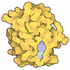
+ Open data
- Basic information
| Entry | Database: PDB / ID: 9jlb | |||||||||||||||||||||
|---|---|---|---|---|---|---|---|---|---|---|---|---|---|---|---|---|---|---|---|---|---|---|
| Title | Cryo-EM structure of phyB-PIF6beta complex | |||||||||||||||||||||
 Components Components |
| |||||||||||||||||||||
 Keywords Keywords | GENE REGULATION / PIF6-mediated / red light / signal transduction / phytochrome B | |||||||||||||||||||||
| Function / homology |  Function and homology information Function and homology informationabscisic acid metabolic process / response to low fluence red light stimulus / red light photoreceptor activity / protein-phytochromobilin linkage / transpiration / red or far-red light signaling pathway / regulation of defense response / far-red light photoreceptor activity / red light signaling pathway / circadian regulation of calcium ion oscillation ...abscisic acid metabolic process / response to low fluence red light stimulus / red light photoreceptor activity / protein-phytochromobilin linkage / transpiration / red or far-red light signaling pathway / regulation of defense response / far-red light photoreceptor activity / red light signaling pathway / circadian regulation of calcium ion oscillation / response to low fluence blue light stimulus by blue low-fluence system / red or far-red light photoreceptor activity / regulation of photoperiodism, flowering / stomatal complex development / gravitropism / phototropism / regulation of seed germination / jasmonic acid mediated signaling pathway / response to far red light / photomorphogenesis / entrainment of circadian clock / response to abscisic acid / detection of visible light / response to salt / response to temperature stimulus / phosphorelay sensor kinase activity / response to light stimulus / photosynthesis / response to cold / promoter-specific chromatin binding / chromatin organization / sequence-specific DNA binding / protein dimerization activity / nuclear speck / nuclear body / DNA-binding transcription factor activity / negative regulation of DNA-templated transcription / protein homodimerization activity / DNA binding / identical protein binding / nucleus / cytosol Similarity search - Function | |||||||||||||||||||||
| Biological species |  | |||||||||||||||||||||
| Method | ELECTRON MICROSCOPY / single particle reconstruction / cryo EM / Resolution: 3 Å | |||||||||||||||||||||
 Authors Authors | Jia, H.L. / Guan, Z.Y. / Ding, J.Y. / Wang, X.Y. / Ma, L. / Yin, P. | |||||||||||||||||||||
| Funding support |  China, 1items China, 1items
| |||||||||||||||||||||
 Citation Citation |  Journal: Cell Discov / Year: 2025 Journal: Cell Discov / Year: 2025Title: Structural insight into PIF6-mediated red light signal transduction of plant phytochrome B. Authors: Hanli Jia / Zeyuan Guan / Junya Ding / Xiaoyu Wang / Dingfang Tian / Yan Zhu / Delin Zhang / Zhu Liu / Ling Ma / Ping Yin /  Abstract: The red/far-red light receptor phytochrome B (phyB) plays essential roles in regulating various plant development processes. PhyB exists in two distinct photoreversible forms: the inactive Pr form ...The red/far-red light receptor phytochrome B (phyB) plays essential roles in regulating various plant development processes. PhyB exists in two distinct photoreversible forms: the inactive Pr form and the active Pfr form. phyB-Pfr binds phytochrome-interacting factors (PIFs) to transduce red light signals. Here, we determined the cryo-electron microscopy (cryo-EM) structures of the photoactivated phyB-Pfr‒PIF6 complex, the constitutively active mutant phyB‒PIF6 complex, and the truncated phyBN‒PIF6 complex. In these structures, two parallel phyB-Pfr molecules interact with one PIF6 molecule. Red light-triggered rotation of the PΦB D-ring leads to the conversion of hairpin loops into α helices and the "head-to-head" reassembly of phyB-Pfr N-terminal photosensory modules. The interaction between phyB-Pfr and PIF6 influences the dimerization and transcriptional activation activity of PIF6, and PIF6 stabilizes the N-terminal extension of phyB-Pfr and increases the Pr→Pfr photoconversion efficiency of phyB. Our findings reveal the molecular mechanisms underlying Pr→Pfr photoconversion and PIF6-mediated red light signal transduction of phyB. | |||||||||||||||||||||
| History |
|
- Structure visualization
| Structure viewer | Molecule:  Molmil Molmil Jmol/JSmol Jmol/JSmol |
|---|
- Downloads & links
- Download
| PDBx/mmCIF format |  9jlb.cif.gz 9jlb.cif.gz | 433.5 KB | Display |  PDBx/mmCIF format PDBx/mmCIF format |
|---|---|---|---|---|
| PDB format |  pdb9jlb.ent.gz pdb9jlb.ent.gz | 335.6 KB | Display |  PDB format PDB format |
| PDBx/mmJSON format |  9jlb.json.gz 9jlb.json.gz | Tree view |  PDBx/mmJSON format PDBx/mmJSON format | |
| Others |  Other downloads Other downloads |
-Validation report
| Summary document |  9jlb_validation.pdf.gz 9jlb_validation.pdf.gz | 571 KB | Display |  wwPDB validaton report wwPDB validaton report |
|---|---|---|---|---|
| Full document |  9jlb_full_validation.pdf.gz 9jlb_full_validation.pdf.gz | 580.1 KB | Display | |
| Data in XML |  9jlb_validation.xml.gz 9jlb_validation.xml.gz | 22.2 KB | Display | |
| Data in CIF |  9jlb_validation.cif.gz 9jlb_validation.cif.gz | 33.5 KB | Display | |
| Arichive directory |  https://data.pdbj.org/pub/pdb/validation_reports/jl/9jlb https://data.pdbj.org/pub/pdb/validation_reports/jl/9jlb ftp://data.pdbj.org/pub/pdb/validation_reports/jl/9jlb ftp://data.pdbj.org/pub/pdb/validation_reports/jl/9jlb | HTTPS FTP |
-Related structure data
| Related structure data |  61582MC  9irkC  9itfC M: map data used to model this data C: citing same article ( |
|---|---|
| Similar structure data | Similarity search - Function & homology  F&H Search F&H Search |
- Links
Links
- Assembly
| Deposited unit | 
|
|---|---|
| 1 |
|
- Components
| #1: Protein | Mass: 135807.547 Da / Num. of mol.: 2 Source method: isolated from a genetically manipulated source Source: (gene. exp.)   #2: Protein | | Mass: 26650.301 Da / Num. of mol.: 1 Source method: isolated from a genetically manipulated source Source: (gene. exp.)   #3: Chemical | Has ligand of interest | Y | Has protein modification | Y | |
|---|
-Experimental details
-Experiment
| Experiment | Method: ELECTRON MICROSCOPY |
|---|---|
| EM experiment | Aggregation state: PARTICLE / 3D reconstruction method: single particle reconstruction |
- Sample preparation
| Component | Name: PhyB(full length)-PIF6beta complex / Type: COMPLEX / Entity ID: #1-#2 / Source: MULTIPLE SOURCES |
|---|---|
| Source (natural) | Organism:  |
| Source (recombinant) | Organism:  |
| Buffer solution | pH: 7.8 |
| Specimen | Conc.: 1 mg/ml / Embedding applied: NO / Shadowing applied: NO / Staining applied: NO / Vitrification applied: YES |
| Vitrification | Cryogen name: ETHANE |
- Electron microscopy imaging
| Experimental equipment |  Model: Tecnai F30 / Image courtesy: FEI Company |
|---|---|
| Microscopy | Model: FEI TECNAI F30 |
| Electron gun | Electron source:  FIELD EMISSION GUN / Accelerating voltage: 300 kV / Illumination mode: FLOOD BEAM FIELD EMISSION GUN / Accelerating voltage: 300 kV / Illumination mode: FLOOD BEAM |
| Electron lens | Mode: BRIGHT FIELD / Nominal defocus max: 1800 nm / Nominal defocus min: 1400 nm |
| Image recording | Electron dose: 50 e/Å2 / Film or detector model: GATAN K3 BIOQUANTUM (6k x 4k) |
- Processing
| EM software | Name: PHENIX / Category: model refinement | ||||||||||||||||||||||||
|---|---|---|---|---|---|---|---|---|---|---|---|---|---|---|---|---|---|---|---|---|---|---|---|---|---|
| CTF correction | Type: NONE | ||||||||||||||||||||||||
| 3D reconstruction | Resolution: 3 Å / Resolution method: FSC 0.143 CUT-OFF / Num. of particles: 429631 / Symmetry type: POINT | ||||||||||||||||||||||||
| Refinement | Highest resolution: 3 Å Stereochemistry target values: REAL-SPACE (WEIGHTED MAP SUM AT ATOM CENTERS) | ||||||||||||||||||||||||
| Refine LS restraints |
|
 Movie
Movie Controller


 PDBj
PDBj


