 Movie
Movie Controller

[English] 日本語
 Yorodumi
- PDB-9jl7: Crystal structure of Actinomycin D-Doxorubicin-d(AGCCGT)2 DNA ter... -
+ Open data

- Basic information
| Entry | Database: PDB / ID: 9jl7 | ||||||
|---|---|---|---|---|---|---|---|
| Title | Crystal structure of Actinomycin D-Doxorubicin-d(AGCCGT)2 DNA ternary complex | ||||||
 Components Components |
| ||||||
 Keywords Keywords | DNA/ANTIBIOTIC / Drug-DNA complex / Actinomycin D / Doxorubicin / DNA / DNA-ANTIBIOTIC complex | ||||||
| Function / homology | Actinomycin D / DOXORUBICIN / : / : / DNA Function and homology information Function and homology information | ||||||
| Biological species | synthetic construct (others) Streptomyces parvulus (bacteria) Streptomyces parvulus (bacteria) | ||||||
| Method |  X-RAY DIFFRACTION / SYNCHROTRON / MOLECULAR REPLACEMENT / Resolution: 1.52 Å X-RAY DIFFRACTION / SYNCHROTRON / MOLECULAR REPLACEMENT / Resolution: 1.52 Å | ||||||
 Authors Authors | Li, H.J. / Satange, R.B. / Hou, M.H. | ||||||
| Funding support |  Taiwan, 1items Taiwan, 1items
| ||||||
 Citation Citation | Journal: J.Med.Chem. / Year: 2025 Title: Structural and Functional Insights into Targeting GCCG Sites in the EGFR Promoter by Two DNA Intercalators to Inhibit Breast Cancer Metastasis. Authors: Chang, C.C. / Li, H.J. / Satange, R. / Lin, S.M. / Chen, T.L. / Lin, C.C. / Neidle, S. / Hou, M.H. | ||||||
| History |
|
- Structure visualization
| Structure viewer | Molecule: MolmilJmol/JSmol |
|---|
- Downloads & links
-Download
| PDBx/mmCIF format | 9jl7.cif.gz | 39.6 KB | Display | PDBx/mmCIF format |
|---|---|---|---|---|
| PDB format | pdb9jl7.ent.gz | 25.9 KB | Display | PDB format |
| PDBx/mmJSON format | 9jl7.json.gz | Tree view | PDBx/mmJSON format | |
| Others |  Other downloads Other downloads |
-Validation report
| Arichive directory | https://data.pdbj.org/pub/pdb/validation_reports/jl/9jl7ftp://data.pdbj.org/pub/pdb/validation_reports/jl/9jl7 | HTTPS FTP |
|---|
-Related structure data
| Similar structure data |
|---|
-Links
 PDBj
PDBj


- Assembly
| Deposited unit | 
| |||||||||||||||
|---|---|---|---|---|---|---|---|---|---|---|---|---|---|---|---|---|
| 1 |
| |||||||||||||||
| Unit cell |
| |||||||||||||||
| Components on special symmetry positions |
|
-Components
-DNA chain , 2 types, 2 molecules AB
| #1: DNA chain | Mass: 1809.217 Da / Num. of mol.: 1 / Source method: obtained synthetically / Source: (synth.) synthetic construct (others) |
|---|---|
| #2: DNA chain | Mass: 1809.217 Da / Num. of mol.: 1 / Source method: obtained synthetically / Source: (synth.) synthetic construct (others) |
-Protein/peptide , 1 types, 1 molecules G
| #3: Protein/peptide | 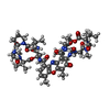  Type: Polypeptide / Class: Antibiotic / Mass: 1291.446 Da / Num. of mol.: 1 / Source method: obtained synthetically Type: Polypeptide / Class: Antibiotic / Mass: 1291.446 Da / Num. of mol.: 1 / Source method: obtained syntheticallyDetails: ACTINOMYCIN D CONSISTS OF TWO PENTAMER RINGS LINKED BY THE CHROMOPHORE (PXZ) Source: (synth.) Streptomyces parvulus (bacteria) / References: NOR: NOR00228, Actinomycin D |
|---|
-Non-polymers , 4 types, 95 molecules 


| #4: Chemical | ChemComp-DM2 /  Mass: 543.519 Da / Num. of mol.: 1 / Source method: obtained synthetically / Formula: C27H29NO11 / Feature type: SUBJECT OF INVESTIGATION / Comment: medication, chemotherapy*YM Mass: 543.519 Da / Num. of mol.: 1 / Source method: obtained synthetically / Formula: C27H29NO11 / Feature type: SUBJECT OF INVESTIGATION / Comment: medication, chemotherapy*YM | ||||
|---|---|---|---|---|---|
| #5: Chemical |  Mass: 54.938 Da / Num. of mol.: 3 / Source method: obtained synthetically / Formula: Mn Mass: 54.938 Da / Num. of mol.: 3 / Source method: obtained synthetically / Formula: Mn#6: Chemical | ChemComp-CL / |  Mass: 35.453 Da / Num. of mol.: 1 / Source method: obtained synthetically / Formula: Cl / Feature type: SUBJECT OF INVESTIGATION Mass: 35.453 Da / Num. of mol.: 1 / Source method: obtained synthetically / Formula: Cl / Feature type: SUBJECT OF INVESTIGATION#7: Water | ChemComp-HOH / | Mass: 18.015 Da / Num. of mol.: 90 / Source method: isolated from a natural source / Formula: H2O |
-Details
| Compound details | ACTINOMYCIN D IS A BICYCLIC PEPTIDE, A MEMBER OF THE ACTINOMYCIN FAMILY. HERE, ACTINOMYCIN D IS ...ACTINOMYCI |
|---|---|
| Has ligand of interest | Y |
| Has protein modification | Y |
-Experimental details
-Experiment
| Experiment | Method: X-RAY DIFFRACTION / Number of used crystals: 1 |
|---|
- Sample preparation
| Crystal | Density Matthews: 2.21 Å3/Da / Density % sol: 44.28 % |
|---|---|
| Crystal grow | Temperature: 293 K / Method: vapor diffusion, sitting drop Details: 0.125mM duplex oligonucleotide, 0.125mM actinomycin D, 0.125mM doxorubicin, 30mM LiCl, 3mM MnCl2, 25mM MES (pH 6.5), 3% PEG 400 |
-Data collection
| Diffraction | Mean temperature: 100 K / Serial crystal experiment: N |
|---|---|
| Diffraction source | Source: SYNCHROTRON / Site: NSRRC / Beamline: BL15A1 / Wavelength: 1 Å |
| Detector | Type: RAYONIX MX300HE / Detector: CCD / Date: Aug 27, 2021 |
| Radiation | Protocol: SINGLE WAVELENGTH / Monochromatic (M) / Laue (L): M / Scattering type: x-ray |
| Radiation wavelength | Wavelength: 1 Å / Relative weight: 1 |
| Reflection | Resolution: 1.52→30 Å / Num. obs: 7813 / % possible obs: 99.7 % / Redundancy: 34.5 % / Rmerge(I) obs: 0.07 / Net I/σ(I): 11.8 |
| Reflection shell | Resolution: 1.52→1.57 Å / Redundancy: 38.8 % / Rmerge(I) obs: 0.449 / Num. unique obs: 729 / CC1/2: 0.994 / CC star: 0.998 / Rpim(I) all: 0.072 / Rrim(I) all: 0.455 / Χ2: 1.096 / % possible all: 100 |
- Processing
| Software |
| |||||||||||||||||||||||||||||||||||||||||||||||||
|---|---|---|---|---|---|---|---|---|---|---|---|---|---|---|---|---|---|---|---|---|---|---|---|---|---|---|---|---|---|---|---|---|---|---|---|---|---|---|---|---|---|---|---|---|---|---|---|---|---|---|
| Refinement | Method to determine structure: MOLECULAR REPLACEMENT / Resolution: 1.52→22.14 Å / SU ML: 0.11 / Cross valid method: NONE / σ(F): 1.34 / Phase error: 21.54 / Stereochemistry target values: ML
| |||||||||||||||||||||||||||||||||||||||||||||||||
| Solvent computation | Shrinkage radii: 0.9 Å / VDW probe radii: 1.11 Å / Solvent model: FLAT BULK SOLVENT MODEL | |||||||||||||||||||||||||||||||||||||||||||||||||
| Refinement step | Cycle: LAST / Resolution: 1.52→22.14 Å
| |||||||||||||||||||||||||||||||||||||||||||||||||
| Refine LS restraints |
| |||||||||||||||||||||||||||||||||||||||||||||||||
| LS refinement shell |
|
