 Movie
Movie Controller

[English] 日本語
 Yorodumi
- PDB-9jjd: Structural analysis of autophagy-related protein 8a in Drosophila... -
+ Open data

- Basic information
| Entry | Database: PDB / ID: 9jjd | ||||||
|---|---|---|---|---|---|---|---|
| Title | Structural analysis of autophagy-related protein 8a in Drosophila melanogaster | ||||||
 Components Components | Autophagy-related 8a, isoform A | ||||||
 Keywords Keywords | LIPID TRANSPORT / LIPID TRAMSPORT / LIPID BINDING PROTEIN | ||||||
| Function / homology |  Function and homology information Function and homology informationTBC/RABGAPs / type I terminal bouton / larval midgut cell programmed cell death / Macroautophagy / amphisome / GABA receptor binding / phosphatidylethanolamine binding / cellular response to nitrogen starvation / reticulophagy / autolysosome ...TBC/RABGAPs / type I terminal bouton / larval midgut cell programmed cell death / Macroautophagy / amphisome / GABA receptor binding / phosphatidylethanolamine binding / cellular response to nitrogen starvation / reticulophagy / autolysosome / autophagosome membrane / regulation of protein ubiquitination / autophagosome maturation / autophagosome assembly / mitophagy / autophagosome / determination of adult lifespan / macroautophagy / autophagy / protein tag activity / ubiquitin protein ligase binding / protein kinase binding Similarity search - Function | ||||||
| Biological species |  | ||||||
| Method |  X-RAY DIFFRACTION / SYNCHROTRON / MOLECULAR REPLACEMENT / Resolution: 1.36 Å X-RAY DIFFRACTION / SYNCHROTRON / MOLECULAR REPLACEMENT / Resolution: 1.36 Å | ||||||
 Authors Authors | Zhang, S.Q. / Luo, X. / Wu, D.X. / Liu, J. / Li, X.Y. | ||||||
| Funding support |  China, 1items China, 1items
| ||||||
 Citation Citation | Journal: J.Agric.Food Chem. / Year: 2025 Title: Crystal Structure of Autophagy-Associated Protein 8 at 1.36 angstrom Resolution and Its Inhibitory Interactions with Indole Analogs. Authors: Zhang, S. / Luo, X. / Yuan, X. / Wu, D. / Liu, J. / Zhao, K. / Xu, Y. / Zhou, J. / Li, X. / Li, Q.X. | ||||||
| History |
|
- Structure visualization
| Structure viewer | Molecule: MolmilJmol/JSmol |
|---|
- Downloads & links
-Download
| PDBx/mmCIF format | 9jjd.cif.gz | 87.2 KB | Display | PDBx/mmCIF format |
|---|---|---|---|---|
| PDB format | pdb9jjd.ent.gz | 66 KB | Display | PDB format |
| PDBx/mmJSON format | 9jjd.json.gz | Tree view | PDBx/mmJSON format | |
| Others |  Other downloads Other downloads |
-Validation report
| Summary document | 9jjd_validation.pdf.gz | 425 KB | Display | wwPDB validaton report |
|---|---|---|---|---|
| Full document | 9jjd_full_validation.pdf.gz | 426.9 KB | Display | |
| Data in XML | 9jjd_validation.xml.gz | 9.4 KB | Display | |
| Data in CIF | 9jjd_validation.cif.gz | 12.2 KB | Display | |
| Arichive directory | https://data.pdbj.org/pub/pdb/validation_reports/jj/9jjdftp://data.pdbj.org/pub/pdb/validation_reports/jj/9jjd | HTTPS FTP |
-Related structure data
| Similar structure data |
|---|
-Links
 PDBj
PDBj
- Assembly
| Deposited unit | 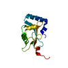
| ||||||||
|---|---|---|---|---|---|---|---|---|---|
| 1 |
| ||||||||
| Unit cell |
|
-Components
| #1: Protein | Mass: 14416.562 Da / Num. of mol.: 1 Source method: isolated from a genetically manipulated source Source: (gene. exp.) Gene: Atg8a, Atg-8a, ATG8, Atg8, atg8, ATG8/LC3, Atg8/LC3, ATG8a, Atg8A, atg8A, atg8a, Atg8alpha, BcDNA:LD05816, CG1534, dAtg8a, DmAtg8a, Dmel\CG32672, DrAtg8a, LC3, LC3/Atg8, CG32672, Dmel_CG32672 Production host:  |
|---|---|
| #2: Water | ChemComp-HOH /  Mass: 18.015 Da / Num. of mol.: 130 / Source method: isolated from a natural source / Formula: H2O Mass: 18.015 Da / Num. of mol.: 130 / Source method: isolated from a natural source / Formula: H2O |
| Has protein modification | N |
-Experimental details
-Experiment
| Experiment | Method: X-RAY DIFFRACTION / Number of used crystals: 1 |
|---|
- Sample preparation
| Crystal | Density Matthews: 2.29 Å3/Da / Density % sol: 46.23 % |
|---|---|
| Crystal grow | Temperature: 293 K / Method: vapor diffusion, hanging drop / Details: 0.1 M Bis-Tris pH=6.5, 26 (W/V) PEG MME 5000 |
-Data collection
| Diffraction | Mean temperature: 100 K / Serial crystal experiment: N |
|---|---|
| Diffraction source | Source: SYNCHROTRON / Site: SSRF / Beamline: BL18U1 / Wavelength: 0.97918 Å |
| Detector | Type: DECTRIS PILATUS3 6M / Detector: PIXEL / Date: Jan 12, 2024 |
| Radiation | Protocol: SINGLE WAVELENGTH / Monochromatic (M) / Laue (L): M / Scattering type: x-ray |
| Radiation wavelength | Wavelength: 0.97918 Å / Relative weight: 1 |
| Reflection | Resolution: 1.36→29.73 Å / Num. obs: 29174 / % possible obs: 99.9 % / Redundancy: 11.7 % / Rmerge(I) obs: 0.085 / Rpim(I) all: 0.025 / Rrim(I) all: 0.085 / Net I/σ(I): 14.7 |
| Reflection shell | Resolution: 1.36→1.4 Å / Rmerge(I) obs: 0.967 / Num. unique obs: 2111 / Rpim(I) all: 0.395 / Rrim(I) all: 1.047 / % possible all: 99.1 |
- Processing
| Software |
| |||||||||||||||||||||||||||||||||||||||||||||||||||||||||||||||||||||||||||||||||||||||||||||||||||||||||
|---|---|---|---|---|---|---|---|---|---|---|---|---|---|---|---|---|---|---|---|---|---|---|---|---|---|---|---|---|---|---|---|---|---|---|---|---|---|---|---|---|---|---|---|---|---|---|---|---|---|---|---|---|---|---|---|---|---|---|---|---|---|---|---|---|---|---|---|---|---|---|---|---|---|---|---|---|---|---|---|---|---|---|---|---|---|---|---|---|---|---|---|---|---|---|---|---|---|---|---|---|---|---|---|---|---|---|
| Refinement | Method to determine structure: MOLECULAR REPLACEMENT / Resolution: 1.36→29.73 Å / SU ML: 0.2 / Cross valid method: THROUGHOUT / σ(F): 1.36 / Phase error: 22.92 / Stereochemistry target values: ML
| |||||||||||||||||||||||||||||||||||||||||||||||||||||||||||||||||||||||||||||||||||||||||||||||||||||||||
| Solvent computation | Shrinkage radii: 0.9 Å / VDW probe radii: 1.1 Å / Solvent model: FLAT BULK SOLVENT MODEL | |||||||||||||||||||||||||||||||||||||||||||||||||||||||||||||||||||||||||||||||||||||||||||||||||||||||||
| Refinement step | Cycle: LAST / Resolution: 1.36→29.73 Å
| |||||||||||||||||||||||||||||||||||||||||||||||||||||||||||||||||||||||||||||||||||||||||||||||||||||||||
| Refine LS restraints |
| |||||||||||||||||||||||||||||||||||||||||||||||||||||||||||||||||||||||||||||||||||||||||||||||||||||||||
| LS refinement shell |
|
