 Movie
Movie Controller

[English] 日本語
 Yorodumi
- PDB-9jgd: Crystal structure of Nep1 in complex with 5'-methylthioadenosine ... -
+ Open data

- Basic information
| Entry | Database: PDB / ID: 9jgd | ||||||
|---|---|---|---|---|---|---|---|
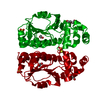
| Title | Crystal structure of Nep1 in complex with 5'-methylthioadenosine from Pyrococcus horikoshii OT3 | ||||||
 Components Components | Ribosomal RNA small subunit methyltransferase Nep1 | ||||||
 Keywords Keywords | TRANSFERASE / Conserved arginine residues / Globular loop extension / Inter-subunit interactions / Nep1 / N1-pseudouridine methyltransferase / Trefoil knot | ||||||
| Function / homology |  Function and homology information Function and homology informationrRNA (pseudouridine) methyltransferase activity / rRNA base methylation / Transferases; Transferring one-carbon groups; Methyltransferases / rRNA binding Similarity search - Function | ||||||
| Biological species |   Pyrococcus horikoshii OT3 (archaea) Pyrococcus horikoshii OT3 (archaea) | ||||||
| Method |  X-RAY DIFFRACTION / MOLECULAR REPLACEMENT / Resolution: 2.2 Å X-RAY DIFFRACTION / MOLECULAR REPLACEMENT / Resolution: 2.2 Å | ||||||
 Authors Authors | Saha, S. / Kanaujia, S.P. | ||||||
| Funding support |  India, 1items India, 1items
| ||||||
 Citation Citation | Journal: Febs J. / Year: 2025 Title: Structural analysis of the ribosome assembly factor Nep1, an N1-specific pseudouridine methyltransferase, reveals mechanistic insights. Authors: Saha, S. / Kanaujia, S.P. | ||||||
| History |
|
- Structure visualization
| Structure viewer | Molecule: MolmilJmol/JSmol |
|---|
- Downloads & links
-Download
| PDBx/mmCIF format | 9jgd.cif.gz | 112.7 KB | Display | PDBx/mmCIF format |
|---|---|---|---|---|
| PDB format | pdb9jgd.ent.gz | 86 KB | Display | PDB format |
| PDBx/mmJSON format | 9jgd.json.gz | Tree view | PDBx/mmJSON format | |
| Others |  Other downloads Other downloads |
-Validation report
| Arichive directory | https://data.pdbj.org/pub/pdb/validation_reports/jg/9jgdftp://data.pdbj.org/pub/pdb/validation_reports/jg/9jgd | HTTPS FTP |
|---|
-Related structure data

-Links
 PDBj
PDBj- Assembly
| Deposited unit | 
| |||||||||
|---|---|---|---|---|---|---|---|---|---|---|
| 1 | 
| |||||||||
| Unit cell |
| |||||||||
| Components on special symmetry positions |
|
-Components
-Protein , 1 types, 1 molecules A
| #1: Protein | Mass: 26926.430 Da / Num. of mol.: 1 Source method: isolated from a genetically manipulated source Source: (gene. exp.) Pyrococcus horikoshii OT3 (archaea) / Gene: nep1, PH1379 / Plasmid: pET28a / Production host:  References: UniProt: O50087, Transferases; Transferring one-carbon groups; Methyltransferases |
|---|
-Non-polymers , 6 types, 96 molecules 


| #2: Chemical | ChemComp-CL /  Mass: 35.453 Da / Num. of mol.: 1 / Source method: obtained synthetically / Formula: Cl Mass: 35.453 Da / Num. of mol.: 1 / Source method: obtained synthetically / Formula: Cl | ||||
|---|---|---|---|---|---|
| #3: Chemical | ChemComp-MTA /  Mass: 297.334 Da / Num. of mol.: 1 / Source method: obtained synthetically / Formula: C11H15N5O3S / Feature type: SUBJECT OF INVESTIGATION Mass: 297.334 Da / Num. of mol.: 1 / Source method: obtained synthetically / Formula: C11H15N5O3S / Feature type: SUBJECT OF INVESTIGATION | ||||
| #4: Chemical | ChemComp-EDO /  Mass: 62.068 Da / Num. of mol.: 1 / Source method: obtained synthetically / Formula: C2H6O2 Mass: 62.068 Da / Num. of mol.: 1 / Source method: obtained synthetically / Formula: C2H6O2 | ||||
| #5: Chemical |  Mass: 92.094 Da / Num. of mol.: 2 / Source method: obtained synthetically / Formula: C3H8O3 Mass: 92.094 Da / Num. of mol.: 2 / Source method: obtained synthetically / Formula: C3H8O3#6: Chemical | ChemComp-ACT / |  Mass: 59.044 Da / Num. of mol.: 1 / Source method: obtained synthetically / Formula: C2H3O2 Mass: 59.044 Da / Num. of mol.: 1 / Source method: obtained synthetically / Formula: C2H3O2#7: Water | ChemComp-HOH / | Mass: 18.015 Da / Num. of mol.: 90 / Source method: isolated from a natural source / Formula: H2O |
-Details
| Has ligand of interest | Y |
|---|---|
| Has protein modification | N |
-Experimental details
-Experiment
| Experiment | Method: X-RAY DIFFRACTION / Number of used crystals: 1 |
|---|
- Sample preparation
| Crystal | Density Matthews: 2.32 Å3/Da / Density % sol: 47 % / Description: Trigonal |
|---|---|
| Crystal grow | Temperature: 293 K / Method: vapor diffusion, hanging drop / pH: 5.6 Details: 0.2M potassium sodium tartrate tetrahydrate, 0.1M sodium citrate tribasic dihydrate pH 5.6, 2.0 ammonium sulfate |
-Data collection
| Diffraction | Mean temperature: 100 K / Serial crystal experiment: N |
|---|---|
| Diffraction source | Source: ROTATING ANODE / Type: RIGAKU MICROMAX-007 HF / Wavelength: 1.5418 Å |
| Detector | Type: RIGAKU RAXIS IV++ / Detector: IMAGE PLATE / Date: Apr 22, 2023 / Details: VariMax HF |
| Radiation | Protocol: SINGLE WAVELENGTH / Monochromatic (M) / Laue (L): M / Scattering type: x-ray |
| Radiation wavelength | Wavelength: 1.5418 Å / Relative weight: 1 |
| Reflection | Resolution: 2.2→70.97 Å / Num. obs: 13053 / % possible obs: 100 % / Redundancy: 12.3 % / CC1/2: 0.995 / Rmerge(I) obs: 0.136 / Rpim(I) all: 0.04 / Rrim(I) all: 0.141 / Χ2: 1.06 / Net I/σ(I): 13.3 / Num. measured all: 160893 |
| Reflection shell | Resolution: 2.2→2.27 Å / % possible obs: 100 % / Redundancy: 12.1 % / Rmerge(I) obs: 0.552 / Num. measured all: 13625 / Num. unique obs: 1127 / CC1/2: 0.952 / Rpim(I) all: 0.164 / Rrim(I) all: 0.576 / Χ2: 1.45 / Net I/σ(I) obs: 5.1 |
- Processing
| Software |
| ||||||||||||||||||||||||||||||||||||||||||||||||||||||||||||||||||||||||||||||||||||||||||||||||||||||||||||||||||||||||||||||||||||||||||||||||||||||||||||||||||||||||||||||||||||||
|---|---|---|---|---|---|---|---|---|---|---|---|---|---|---|---|---|---|---|---|---|---|---|---|---|---|---|---|---|---|---|---|---|---|---|---|---|---|---|---|---|---|---|---|---|---|---|---|---|---|---|---|---|---|---|---|---|---|---|---|---|---|---|---|---|---|---|---|---|---|---|---|---|---|---|---|---|---|---|---|---|---|---|---|---|---|---|---|---|---|---|---|---|---|---|---|---|---|---|---|---|---|---|---|---|---|---|---|---|---|---|---|---|---|---|---|---|---|---|---|---|---|---|---|---|---|---|---|---|---|---|---|---|---|---|---|---|---|---|---|---|---|---|---|---|---|---|---|---|---|---|---|---|---|---|---|---|---|---|---|---|---|---|---|---|---|---|---|---|---|---|---|---|---|---|---|---|---|---|---|---|---|---|---|
| Refinement | Method to determine structure: MOLECULAR REPLACEMENT / Resolution: 2.2→70.97 Å / Cor.coef. Fo:Fc: 0.955 / Cor.coef. Fo:Fc free: 0.926 / SU B: 15.044 / SU ML: 0.183 / Cross valid method: THROUGHOUT / ESU R: 0.285 / ESU R Free: 0.226 / Stereochemistry target values: MAXIMUM LIKELIHOOD / Details: HYDROGENS HAVE BEEN ADDED IN THE RIDING POSITIONS
| ||||||||||||||||||||||||||||||||||||||||||||||||||||||||||||||||||||||||||||||||||||||||||||||||||||||||||||||||||||||||||||||||||||||||||||||||||||||||||||||||||||||||||||||||||||||
| Solvent computation | Ion probe radii: 0.8 Å / Shrinkage radii: 0.8 Å / VDW probe radii: 1.2 Å / Solvent model: MASK | ||||||||||||||||||||||||||||||||||||||||||||||||||||||||||||||||||||||||||||||||||||||||||||||||||||||||||||||||||||||||||||||||||||||||||||||||||||||||||||||||||||||||||||||||||||||
| Displacement parameters | Biso mean: 41.882 Å2
| ||||||||||||||||||||||||||||||||||||||||||||||||||||||||||||||||||||||||||||||||||||||||||||||||||||||||||||||||||||||||||||||||||||||||||||||||||||||||||||||||||||||||||||||||||||||
| Refinement step | Cycle: 1 / Resolution: 2.2→70.97 Å
| ||||||||||||||||||||||||||||||||||||||||||||||||||||||||||||||||||||||||||||||||||||||||||||||||||||||||||||||||||||||||||||||||||||||||||||||||||||||||||||||||||||||||||||||||||||||
| Refine LS restraints |
|