 Movie
Movie Controller


+ Open data

- Basic information
| Entry | Database: PDB / ID: 9jg6 | ||||||||||||||||||||||||
|---|---|---|---|---|---|---|---|---|---|---|---|---|---|---|---|---|---|---|---|---|---|---|---|---|---|
| Title | The tail-complex structure of phage P22 | ||||||||||||||||||||||||
 Components Components |
| ||||||||||||||||||||||||
 Keywords Keywords | VIRAL PROTEIN / Complex | ||||||||||||||||||||||||
| Function / homology |  Function and homology information Function and homology informationBacteriophage P22, Gp10, DNA-stabilising / Phage stabilisation protein / Tail accessory factor GP4 / Peptidoglycan hydrolase Gp4 superfamily / P22 tail accessory factor / Phage P22-like portal protein / Phage P22-like portal protein / P22 tailspike C-terminal domain / Salmonella phage P22 tail-spike / Bacteriophage P22 tailspike, N-terminal ...Bacteriophage P22, Gp10, DNA-stabilising / Phage stabilisation protein / Tail accessory factor GP4 / Peptidoglycan hydrolase Gp4 superfamily / P22 tail accessory factor / Phage P22-like portal protein / Phage P22-like portal protein / P22 tailspike C-terminal domain / Salmonella phage P22 tail-spike / Bacteriophage P22 tailspike, N-terminal / Phage P22 tailspike-like, N-terminal domain superfamily / Head binding / Autotransporter, pectate lyase C-like domain superfamily / Pectin lyase fold/virulence factor Similarity search - Domain/homology | ||||||||||||||||||||||||
| Biological species |  Salmonella enterica subsp. enterica serovar Typhimurium (bacteria) Salmonella enterica subsp. enterica serovar Typhimurium (bacteria) | ||||||||||||||||||||||||
| Method | ELECTRON MICROSCOPY / single particle reconstruction / cryo EM / Resolution: 3.21 Å | ||||||||||||||||||||||||
 Authors Authors | Liu, H.R. / Xiao, H. | ||||||||||||||||||||||||
| Funding support |  China, 3items China, 3items
| ||||||||||||||||||||||||
 Citation Citation | Journal: PLoS Biol / Year: 2025 Title: Structure of the scaffolding protein and portal within the bacteriophage P22 procapsid provides insights into the self-assembly process. Authors: Hao Xiao / Wenyuan Chen / Hao Pang / Jing Zheng / Li Wang / Hao Feng / Jingdong Song / Lingpeng Cheng / Hongrong Liu / Abstract: In the assembly pathway of tailed double-stranded DNA (dsDNA) bacteriophages and herpesviruses, a procapsid with a dodecameric portal for DNA delivery at a unique vertex is initially formed. ...In the assembly pathway of tailed double-stranded DNA (dsDNA) bacteriophages and herpesviruses, a procapsid with a dodecameric portal for DNA delivery at a unique vertex is initially formed. Appropriate procapsid assembly requires the transient presence of multiple copies of a scaffolding protein (SP), which is absent in the mature virion. However, how the SP contributes to dodecameric portal formation, facilitates portal and coat protein incorporation, and is subsequently released remains unclear because of a lack of structural information. Here, we present the structure of the SP-portal complex within the procapsid of bacteriophage P22 at 3-9 Å resolutions. The AlphaFold2-predicted SP model fits well with the density map of the complex. The SP forms trimers and tetramers that interact to yield a dome-like complex on the portal. Two SP domains mediate multimerization. Each trimer interacts with two neighboring portal subunits. The SP has a loop-hook-like structure that aids in coat protein recruitment during viral assembly. The loops of those SP subunits on the portal are positioned in clefts between adjacent portal subunits. Conformational changes in the portal during phage maturation may trigger the disassembly and release of the SP complex. Our findings provide insights into SP-assisted procapsid assembly in bacteriophage P22 and suggest that this strategy is also implemented by other dsDNA viruses, including herpesviruses. | ||||||||||||||||||||||||
| History |
|
- Structure visualization
| Structure viewer | Molecule: MolmilJmol/JSmol |
|---|
- Downloads & links
-Download
| PDBx/mmCIF format | 9jg6.cif.gz | 2.5 MB | Display | PDBx/mmCIF format |
|---|---|---|---|---|
| PDB format | pdb9jg6.ent.gz | Display | PDB format | |
| PDBx/mmJSON format | 9jg6.json.gz | Tree view | PDBx/mmJSON format | |
| Others |  Other downloads Other downloads |
-Validation report
| Summary document | 9jg6_validation.pdf.gz | 1.6 MB | Display | wwPDB validaton report |
|---|---|---|---|---|
| Full document | 9jg6_full_validation.pdf.gz | 1.6 MB | Display | |
| Data in XML | 9jg6_validation.xml.gz | 311.3 KB | Display | |
| Data in CIF | 9jg6_validation.cif.gz | 499.5 KB | Display | |
| Arichive directory | https://data.pdbj.org/pub/pdb/validation_reports/jg/9jg6ftp://data.pdbj.org/pub/pdb/validation_reports/jg/9jg6 | HTTPS FTP |
-Related structure data
| Related structure data | 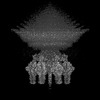 61457MC  9jgaC  9kyvC  9kywC  9kyxC  9kyyC M: map data used to model this data C: citing same article ( |
|---|---|
| Similar structure data |


-Links
 PDBj
PDBj- Assembly
| Deposited unit | 
|
|---|---|
| 1 |
|
-Components
| #1: Protein | Mass: 82829.375 Da / Num. of mol.: 12 / Source method: isolated from a natural source Source: (natural) Salmonella enterica subsp. enterica serovar Typhimurium (bacteria)References: UniProt: A0A3V9J0D3 #2: Protein | Mass: 71923.523 Da / Num. of mol.: 18 / Source method: isolated from a natural source Source: (natural) Salmonella enterica subsp. enterica serovar Typhimurium (bacteria)References: UniProt: A0A3V9J050 #3: Protein | Mass: 52512.020 Da / Num. of mol.: 6 / Source method: isolated from a natural source Source: (natural) Salmonella enterica subsp. enterica serovar Typhimurium (bacteria)References: UniProt: A0A444A1G7 #4: Protein | Mass: 18044.959 Da / Num. of mol.: 12 / Source method: isolated from a natural source Source: (natural) Salmonella enterica subsp. enterica serovar Typhimurium (bacteria)References: UniProt: A0A444A265 Has protein modification | N | |
|---|
-Experimental details
-Experiment
| Experiment | Method: ELECTRON MICROSCOPY |
|---|---|
| EM experiment | Aggregation state: PARTICLE / 3D reconstruction method: single particle reconstruction |
- Sample preparation
| Component | Name: Salmonella phage P22 / Type: COMPLEX / Entity ID: all / Source: NATURAL |
|---|---|
| Source (natural) | Organism: Salmonella enterica subsp. enterica serovar Typhimurium (bacteria) |
| Details of virus | Empty: NO / Enveloped: NO / Isolate: SPECIES / Type: VIRION |
| Buffer solution | pH: 7.4 |
| Specimen | Embedding applied: NO / Shadowing applied: NO / Staining applied: NO / Vitrification applied: YES |
| Vitrification | Cryogen name: ETHANE |
- Electron microscopy imaging
| Experimental equipment |  Model: Titan Krios / Image courtesy: FEI Company |
|---|---|
| Microscopy | Model: TFS KRIOS |
| Electron gun | Electron source:  FIELD EMISSION GUN / Accelerating voltage: 300 kV / Illumination mode: FLOOD BEAM FIELD EMISSION GUN / Accelerating voltage: 300 kV / Illumination mode: FLOOD BEAM |
| Electron lens | Mode: BRIGHT FIELD / Nominal defocus max: 2200 nm / Nominal defocus min: 1600 nm |
| Image recording | Electron dose: 32 e/Å2 / Film or detector model: GATAN K3 (6k x 4k) |
- Processing
| CTF correction | Type: PHASE FLIPPING ONLY |
|---|---|
| 3D reconstruction | Resolution: 3.21 Å / Resolution method: FSC 0.143 CUT-OFF / Num. of particles: 82501 / Symmetry type: POINT |
