 Movie
Movie Controller

[English] 日本語
 Yorodumi
- PDB-9jfc: Crystal structure of Pseudomonas aeruginosa SuhB complexed with G... -
+ Open data

- Basic information
| Entry | Database: PDB / ID: 9jfc | ||||||
|---|---|---|---|---|---|---|---|
| Title | Crystal structure of Pseudomonas aeruginosa SuhB complexed with Gallic acid in monoclinic space group | ||||||
 Components Components | Nus factor SuhB | ||||||
 Keywords Keywords | HYDROLASE / Phytochemical | ||||||
| Function / homology |  Function and homology information Function and homology informationinositol-phosphate phosphatase / inositol monophosphate 1-phosphatase activity / inositol metabolic process / phosphatidylinositol phosphate biosynthetic process / transcription antitermination / ribosome biogenesis / DNA-templated transcription / signal transduction / RNA binding / metal ion binding / cytoplasm Similarity search - Function | ||||||
| Biological species |   Pseudomonas aeruginosa (bacteria) Pseudomonas aeruginosa (bacteria) | ||||||
| Method |  X-RAY DIFFRACTION / SYNCHROTRON / MOLECULAR REPLACEMENT / Resolution: 2.2 Å X-RAY DIFFRACTION / SYNCHROTRON / MOLECULAR REPLACEMENT / Resolution: 2.2 Å | ||||||
 Authors Authors | Yadav, V.K. / Shukla, M. / Maji, S. / Bhattacharyya, S. | ||||||
| Funding support |  India, 1items India, 1items
| ||||||
 Citation Citation | Journal: To Be Published Title: Crystal structure of Pseudomonas aeruginosa SuhB complexed with Gallic acid in monoclinic space group Authors: Yadav, V.K. / Shukla, M. / Maji, S. / Bhattacharyya, S. | ||||||
| History |
|
- Structure visualization
| Structure viewer | Molecule: MolmilJmol/JSmol |
|---|
- Downloads & links
-Download
| PDBx/mmCIF format | 9jfc.cif.gz | 416.7 KB | Display | PDBx/mmCIF format |
|---|---|---|---|---|
| PDB format | pdb9jfc.ent.gz | 342.9 KB | Display | PDB format |
| PDBx/mmJSON format | 9jfc.json.gz | Tree view | PDBx/mmJSON format | |
| Others |  Other downloads Other downloads |
-Validation report
| Arichive directory | https://data.pdbj.org/pub/pdb/validation_reports/jf/9jfcftp://data.pdbj.org/pub/pdb/validation_reports/jf/9jfc | HTTPS FTP |
|---|
-Related structure data
| Similar structure data |
|---|
-Links
 PDBj
PDBj


- Assembly
| Deposited unit | 
| |||||||||||||||||||||
|---|---|---|---|---|---|---|---|---|---|---|---|---|---|---|---|---|---|---|---|---|---|---|
| 1 | 
| |||||||||||||||||||||
| 2 | 
| |||||||||||||||||||||
| Unit cell |
| |||||||||||||||||||||
| Noncrystallographic symmetry (NCS) | NCS domain:
|
-Components
| #1: Protein | Mass: 29668.654 Da / Num. of mol.: 4 Source method: isolated from a genetically manipulated source Details: The starting two amino acids are cloning artifacts (coming from the plasmid vector). Source: (gene. exp.) Pseudomonas aeruginosa (bacteria) / Strain: PAO1 / Gene: suhB, PA3818 / Production host: #2: Chemical | ChemComp-GOL /   Mass: 92.094 Da / Num. of mol.: 5 / Source method: obtained synthetically / Formula: C3H8O3 Mass: 92.094 Da / Num. of mol.: 5 / Source method: obtained synthetically / Formula: C3H8O3#3: Chemical | ChemComp-GDE / 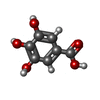  Mass: 170.120 Da / Num. of mol.: 4 / Source method: obtained synthetically / Formula: C7H6O5 / Feature type: SUBJECT OF INVESTIGATION Mass: 170.120 Da / Num. of mol.: 4 / Source method: obtained synthetically / Formula: C7H6O5 / Feature type: SUBJECT OF INVESTIGATION#4: Chemical |   Mass: 154.251 Da / Num. of mol.: 2 / Source method: obtained synthetically / Formula: C4H10O2S2 Mass: 154.251 Da / Num. of mol.: 2 / Source method: obtained synthetically / Formula: C4H10O2S2#5: Water | ChemComp-HOH / |  Mass: 18.015 Da / Num. of mol.: 333 / Source method: isolated from a natural source / Formula: H2O Mass: 18.015 Da / Num. of mol.: 333 / Source method: isolated from a natural source / Formula: H2OHas ligand of interest | Y | Has protein modification | N | |
|---|
-Experimental details
-Experiment
| Experiment | Method: X-RAY DIFFRACTION / Number of used crystals: 1 |
|---|
- Sample preparation
| Crystal | Density Matthews: 2.14 Å3/Da / Density % sol: 42.53 % |
|---|---|
| Crystal grow | Temperature: 298 K / Method: vapor diffusion, hanging drop / Details: PEG 3350, Sodium acetate trihydrate |
-Data collection
| Diffraction | Mean temperature: 100 K / Serial crystal experiment: N |
|---|---|
| Diffraction source | Source: SYNCHROTRON / Site: RRCAT INDUS-2 / Beamline: PX-BL21 / Wavelength: 0.97893 Å |
| Detector | Type: MAR scanner 345 mm plate / Detector: IMAGE PLATE / Date: Jun 24, 2024 |
| Radiation | Protocol: SINGLE WAVELENGTH / Monochromatic (M) / Laue (L): M / Scattering type: x-ray |
| Radiation wavelength | Wavelength: 0.97893 Å / Relative weight: 1 |
| Reflection | Resolution: 2.2→45.3 Å / Num. obs: 50522 / % possible obs: 99.6 % / Redundancy: 3.8 % / CC1/2: 0.995 / Rmerge(I) obs: 0.089 / Rpim(I) all: 0.054 / Rrim(I) all: 0.104 / Net I/σ(I): 10.5 / Num. measured all: 189644 |
| Reflection shell | Resolution: 2.2→2.32 Å / % possible obs: 99.9 % / Redundancy: 3.8 % / Rmerge(I) obs: 0.282 / Num. measured all: 27802 / Num. unique obs: 7363 / CC1/2: 0.928 / Rpim(I) all: 0.168 / Rrim(I) all: 0.329 / Net I/σ(I) obs: 4.5 |
- Processing
| Software |
| ||||||||||||||||||||||||||||||||||||||||||||||||||||||||||||||||||||||||||||||||||||||||||||||||||||||||||||||||||||||||||||||||||||||||||||||||||||||||||||||||||||||||||||||||||||||
|---|---|---|---|---|---|---|---|---|---|---|---|---|---|---|---|---|---|---|---|---|---|---|---|---|---|---|---|---|---|---|---|---|---|---|---|---|---|---|---|---|---|---|---|---|---|---|---|---|---|---|---|---|---|---|---|---|---|---|---|---|---|---|---|---|---|---|---|---|---|---|---|---|---|---|---|---|---|---|---|---|---|---|---|---|---|---|---|---|---|---|---|---|---|---|---|---|---|---|---|---|---|---|---|---|---|---|---|---|---|---|---|---|---|---|---|---|---|---|---|---|---|---|---|---|---|---|---|---|---|---|---|---|---|---|---|---|---|---|---|---|---|---|---|---|---|---|---|---|---|---|---|---|---|---|---|---|---|---|---|---|---|---|---|---|---|---|---|---|---|---|---|---|---|---|---|---|---|---|---|---|---|---|---|
| Refinement | Method to determine structure: MOLECULAR REPLACEMENT / Resolution: 2.2→44.98 Å / Cor.coef. Fo:Fc: 0.946 / Cor.coef. Fo:Fc free: 0.908 / SU B: 8.198 / SU ML: 0.113 / Cross valid method: THROUGHOUT / ESU R: 0.071 / ESU R Free: 0.047 / Stereochemistry target values: MAXIMUM LIKELIHOOD / Details: HYDROGENS HAVE BEEN ADDED IN THE RIDING POSITIONS
| ||||||||||||||||||||||||||||||||||||||||||||||||||||||||||||||||||||||||||||||||||||||||||||||||||||||||||||||||||||||||||||||||||||||||||||||||||||||||||||||||||||||||||||||||||||||
| Solvent computation | Ion probe radii: 0.8 Å / Shrinkage radii: 0.8 Å / VDW probe radii: 1.2 Å / Solvent model: MASK | ||||||||||||||||||||||||||||||||||||||||||||||||||||||||||||||||||||||||||||||||||||||||||||||||||||||||||||||||||||||||||||||||||||||||||||||||||||||||||||||||||||||||||||||||||||||
| Displacement parameters | Biso mean: 24.758 Å2
| ||||||||||||||||||||||||||||||||||||||||||||||||||||||||||||||||||||||||||||||||||||||||||||||||||||||||||||||||||||||||||||||||||||||||||||||||||||||||||||||||||||||||||||||||||||||
| Refinement step | Cycle: 1 / Resolution: 2.2→44.98 Å
| ||||||||||||||||||||||||||||||||||||||||||||||||||||||||||||||||||||||||||||||||||||||||||||||||||||||||||||||||||||||||||||||||||||||||||||||||||||||||||||||||||||||||||||||||||||||
| Refine LS restraints |
|